
Medical expert of the article

New publications
Optical coherence tomography of the eye: what the test shows, when it is prescribed, and how to correctly interpret the results
Last updated: 18.03.2026

All iLive content is medically reviewed or fact checked to ensure as much factual accuracy as possible.
We have strict sourcing guidelines and only link to reputable media sites, academic research institutions and, whenever possible, medically peer reviewed studies. Note that the numbers in parentheses ([1], [2], etc.) are clickable links to these studies.
If you feel that any of our content is inaccurate, out-of-date, or otherwise questionable, please select it and press Ctrl + Enter.
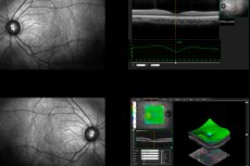
Optical coherence tomography is a non-invasive imaging technique that uses light to produce cross-sectional images of ocular tissue. In ophthalmology practice, it allows for rapid visualization of the retina, macula, optic nerve, and, in some modes, the anterior segment of the eye, without touching the surface of the eye with an instrument. This is why the technique has become routine not only in large specialized centers but also in routine clinical work. [1]
The historical significance of this method is difficult to overstate. The US National Eye Institute directly attributes the modern era of optical coherence tomography to the work of James Fujimoto, David Huang, and Eric Swanson, while the Proceedings of the National Academy of Sciences and the Journal of the American Medical Association consider the invention of this method to be an event that radically changed the diagnosis of eye diseases. This recognition was recognized by the Lasker Award, presented to its developers in 2023. [2]
Today, the method is no longer perceived as a pretty picture. Clinical reviews and educational sources describe it as the preferred imaging method for macular pathology and an important monitoring tool for glaucoma, and modern reviews emphasize its routine use in retinal diseases, including age-related macular degeneration and diabetic retinopathy. [3]
The method is especially useful when it's important for the physician to not just detect a disease but also to see its microstructure. In age-related macular degeneration, layer-by-layer imaging helps identify subretinal and intraretinal fluid, and in diabetic retinal disease, the method is used to monitor macular edema and treatment effectiveness. Several recent reviews of clinical guidelines indicate that optical coherence tomography has become central to the management of neovascular age-related macular degeneration. [4]
Although in everyday speech this method is most often associated with the eyes, its significance is broader. Optical coherence tomography is also used in cardiology as an intravascular imaging method, for example, for navigation during percutaneous coronary interventions. This underscores the general principle of the technology: it provides the physician with a highly detailed image of tissue in areas where conventional imaging methods are no longer sufficient. [5]
Below is a brief summary of what exactly the doctor gets with the method. [6]
| What is being assessed? | What does this give to the doctor? | Where it is especially important |
|---|---|---|
| Makulu | Thickness, presence of fluid, traction, defects | Age-related macular degeneration, macular edema, macular hole |
| Retina by layers | Structural changes at the microscopic level | Diabetic retinopathy, retinal vascular diseases |
| Optic nerve and nerve fiber layer | Signs of thinning and progression | Glaucoma and suspected glaucoma |
| Anterior segment of the eye | Corneal configuration, anterior chamber angle, postoperative changes | Trauma, keratitis, surgical planning |
| Vascular networks without dye | Microcirculation in the retina and choroid | Diabetic retinopathy, vascular and neuro-ophthalmological conditions |
The content of the table is based on clinical reviews and guidelines for the application of the method in retinal pathology, glaucoma and the anterior segment of the eye. [7]
How the method works and which of its varieties are really important
The physical basis of the method is low-coherence optical interferometry. Simply put, the device analyzes reflected light and, using this signal, constructs a layer-by-layer tissue section. The U.S. Food and Drug Administration classifies such devices as devices for viewing, measuring, and analyzing ocular structures, and their technical method is explicitly described as obtaining transverse tomograms of ocular tissue using a non-invasive, contactless method. [8]
Technology has evolved from earlier time-domain systems to more modern spectral-domain systems. A current review from 2025 emphasizes that new spectral-domain devices use a broadband light source, provide higher axial resolution, capture images faster, reduce motion artifacts, and allow for the acquisition of a series of sequential sections in a short time. This is important not only for the image's aesthetic appeal but also for its diagnostic reliability. [9]
The next step is 3D reconstruction. From multiple 2D slices, the device creates a three-dimensional image of the retina, calculates tissue thickness and volume, and allows for comparisons between examination dates. In the clinic, this is especially useful when assessing progress: whether swelling is decreasing, whether nerve fiber thinning is progressing, or whether the process has stabilized after treatment. [10]
A separate area is angiographic optical coherence tomography. It reveals the retinal and choroidal vascular networks without the introduction of dye and has already changed the approach to studying microcirculation. However, reviews from 2024 and 2025 emphasize that interpreting vascular images remains complex: findings are not always specific, different devices do not provide fully comparable results, and standard comparison bases are still far from ideal. [11]
Other important advances include wide-field imaging, portable handheld systems, intraoperative solutions, and home monitoring. Recent publications describe the expansion of the field of view in wide-field imaging, the clinical value of handheld and operative systems, and the emergence of remote monitoring and research devices aimed at more accessible screening. All of this suggests that the technology has long since moved beyond the standard diagnostic room. [12]
Below is a practical table of the main types of the method. [13]
| Variety | The main task | Strong point | Limitation |
|---|---|---|---|
| Temporary | Basic Structural Visualization | A historically important stage of development | Lower speed and detail |
| Spectral-domain | Everyday structural diagnostics | Fast scanning, good resolution | Sensitive to motion artifacts and haze |
| With a swept source | Deeper and broader visualization | Deep structures and periphery are better visible | Not available everywhere |
| Angiographic | Dye-free microcirculation assessment | Layered vascular map | Artifacts, inter-device variability |
| Anterior segment | Evaluation of the cornea, angle, and postoperative changes | Useful in trauma and surgery | Does not replace a full clinical examination |
| Home | Observation between visits | Frequent remote monitoring in selected patients | It does not replace standard treatment and is not intended for independent treatment decisions. |
The table is based on regulatory materials, current technology reviews, and clinical application publications.[14]
When is a study really necessary and when is it just a formality?
The most typical indication is diseases of the macula and retina. The method is particularly valuable in age-related macular degeneration, diabetic macular edema, retinal vein occlusions, epiretinal membranes, and macular holes, as it allows for the visualization of fluid, traction, thickening, and microstructural deformations that are much more crudely assessed by a standard fundus examination. Modern reviews clearly indicate the routine use of the method in age-related macular degeneration and diabetic retinopathy. [15]
The second major group of indications is glaucoma and suspected glaucoma. Here, the method helps evaluate the retinal nerve fiber layer, the neuroretinal rim, and the ganglion complex in the macula. Recent publications emphasize that optical coherence tomography has become a valuable adjunct to perimetry in monitoring glaucoma, as it reveals morphological changes even before the patient notices significant visual impairment. [16]
Diabetic retinopathy occupies a special place. In 2024, the UK National Institute for Health and Care Excellence (NIH) published a special review of the accuracy of ultra-widefield fundus photography and optical coherence tomography for monitoring diabetic retinopathy and diabetic macular edema, which in itself demonstrates how deeply this method is integrated into the modern monitoring pathway. Furthermore, publications of the American Academy of Ophthalmology and reviews of widefield angiographic tomography emphasize its value in detecting early signs and assessing the severity of vascular changes. [17]
The method is increasingly being used in the anterior segment of the eye. Current reviews demonstrate its usefulness in cases of trauma, corneal foreign bodies, infectious keratitis, postoperative monitoring, assessment of filtration pads, and intervention planning. For the physician, it is especially useful in cases where not only an external examination is needed but also an understanding of the depth, width, configuration, and layered structure of the lesion. [18]
Finally, there is growing evidence about the method's role in systems medicine. The angiographic version allows for the visualization of microvascular changes associated with diabetes, hypertension, cardiovascular, and neurodegenerative conditions, while intravascular optical coherence tomography is used in cardiology for navigation during coronary artery interventions. In other words, this method is increasingly being viewed as a platform for high-precision imaging, and not just as a narrowly ophthalmological examination. [19]
Below is a table that helps you quickly understand when a study provides real clinical benefit. [20]
| Situation | Why is it prescribed? | What are they looking for? |
|---|---|---|
| Decreased central vision | Clarify the condition of the macula | Swelling, fluid, membranes, hole |
| Suspected glaucoma | Assess the structure of the optic nerve | Thinning of the nerve fiber layer and ganglion complex |
| Diabetes | Monitoring retinal damage and macular edema | Thickening, cysts, vascular changes |
| Age-related macular degeneration | Confirm the activity of the process and observe it dynamically | Subretinal and intraretinal fluid, pigment epithelial detachment |
| Trauma or keratitis | Specify the depth and configuration of the lesion | Corneal defects, foreign bodies, anterior segment changes |
| Cardiological intervention | To clarify the morphology of the vascular wall | The structure of the plaque, stent, and vessel lumen |
The table is compiled based on clinical reviews of the use of the method in ophthalmology and cardiology. [21]
How the test is performed and how the doctor usually reads the results
For the patient, the examination is usually straightforward. In most cases, no special preparation is required, but sometimes the doctor dilates the pupil with drops, especially if the back of the eye needs to be examined better. After this, the patient is asked to sit in front of the machine, rest their chin on the support, and look at the light marker. The imaging itself is short and non-invasive. [22]
It's important to understand that after pupil dilation, blurred vision and light sensitivity may temporarily occur. Some hospital instructions specifically state that driving is not recommended during these hours. The scan itself is painless and does not require the device to come into contact with the eye, so any discomfort is most often related to the drops, if used, rather than the tomography itself. [23]
The result is not a single image, but a complete data set. The physician receives a series of cross-sections, a thickness map, sometimes a 3D reconstruction, and, in angiographic mode, a layered map of the vessels. Modern reviews emphasize that it is the combination of high-quality images and quantitative parameters that makes the method particularly suitable for long-term observation. [24]
When interpreting a structural examination, the physician looks at the shape of the foveal pit, the integrity of the retinal layers, the thickness of the central zone, the presence of intraretinal and subretinal fluid, epiretinal membrane, vitreomacular traction, pigment epithelial detachment, or signs of atrophy. In glaucoma, the emphasis shifts to the nerve fiber layer, neuroretinal rim, and macular ganglion complex. Therefore, the same device can be used for completely different clinical applications. [25]
One of the most common mistakes patients make is to perceive the thickness number as a definitive diagnosis. In reality, the doctor almost always evaluates not only the number, but also the shape of the layers, the clinical picture, complaints, examination data, previous imaging studies, and the results of other methods. In glaucoma, for example, it's not a single measurement that's particularly important, but rather an analysis of events and trends over time, as clearly demonstrated by modern reviews of disease progression. [26]
Below is a simplified table of the logic of interpretation. [27]
| What does the doctor see? | Possible meaning | Where is it most common? |
|---|---|---|
| Preserved layering and smooth contour of the macula | Close to normal picture | Absence of active macular pathology |
| Intraretinal cysts | Macular edema | Diabetes, retinal vascular diseases |
| Subretinal fluid | Activity of the neovascular process | Age-related macular degeneration |
| Epiretinal membrane and traction | Mechanical deformation of the macula | Membranes, vitreomacular syndrome |
| Thinning of the nerve fiber layer | Structural lesion of the optic nerve | Glaucoma |
| An unusual vascular map without dye | Microcirculation disorder | Diabetic retinopathy, vascular and systemic conditions |
The table reflects a general approach to interpretation, but the final conclusion always depends on the clinical context and the dynamics of observation. [28]
Strengths of the method, its limitations and causes of diagnostic errors
The main advantage of this method is its combination of high detail, non-invasiveness, and repeatability. It provides the physician with objective images and quantitative data that can be compared over time, and allows the patient to undergo the examination repeatedly without the burden associated with invasive procedures. This is precisely why the method is so convenient for long-term monitoring of chronic eye diseases. [29]
But the method also has important limitations. It does not replace a full ophthalmological examination, does not replace fundus examination, perimetry, or dye angiography where necessary, and, most importantly, should not be interpreted separately from complaints and clinical presentation. Even home tomography versions, which have become available in recent years, are officially positioned as a monitoring tool between visits, not as a replacement for a standard examination or as a means of making independent treatment decisions. [30]
Particular caution is required with angiographic studies. A large review published in 2024 highlights several issues: the relative nonspecificity of findings, differences between devices, the lack of a fully standardized regulatory framework, and the presence of artifacts that can distort the vascular image. Therefore, a "beautiful" image of the vessels does not automatically mean an accurate diagnosis. [31]
Artifacts are not uncommon, but a real practical problem. In a 2024 study, only 9.4% of images with formally acceptable signal strength were completely free of artifacts; the most common were shadows, which often obscured critical areas of the image. For the clinician, this implies a simple rule: a good quality score does not guarantee an error-free image. [32]
There are also clinical limitations. Clouding of the optical media, poor visual fixation, severe dry eye syndrome, patient movement, incorrect layer segmentation by the software, high myopia, severe cicatricial changes, and severe edema can significantly impair the quality of the examination. Therefore, interpretation always requires experience, and any questionable results should be reconfirmed either by repeat scanning or other methods. [33]
Below is a table that helps to understand the strengths and weaknesses of the method without oversimplifying. [34]
| Parameter | Strong point | Limitation |
|---|---|---|
| Safety | Non-contact examination, suitable for repeat examinations | When the pupil dilates, temporary discomfort and blurred vision may occur. |
| Detailing | Layered changes are visible that are not noticeable during a normal examination. | Not all structural changes clearly indicate one disease |
| Dynamics | It is convenient to compare studies over time | Comparability depends on quality and identical protocols |
| Angiographic mode | Vessels are visible without the introduction of dye | Many artifacts and inter-device differences |
| Home monitoring | Allows for more frequent monitoring of individual patients | It does not replace an in-person examination and is not intended for independent treatment selection. |
| Automatic calculations | Quickly produce numerical indicators | Segmentation errors and a false sense of accuracy are possible |
The table is based on regulatory documents, reviews of the angiographic version of the method and artifact studies. [35]
New Directions: What's Changing Right Now
One of the most notable trends in recent years is home monitoring. In May 2024, the U.S. Food and Drug Administration issued a designation for a new category of home ophthalmic optical coherence tomography systems for monitoring. This is an important step toward more frequent monitoring of patients with neovascular age-related macular degeneration between scheduled visits. [36]
At the same time, the regulatory logic remains very cautious. The decision materials emphasize that the home system is intended for recording between standard clinical examinations and should not replace in-person visits or be used for independent treatment decisions. In other words, the approach is promising, but not magical: it expands monitoring, not replaces the doctor. [37]
The second trend is strengthening the analytical component of the method. A 2024 review of the angiographic version specifically highlights quantitative image analysis, combination with artificial intelligence, and multimodal visualization as key development vectors. Publications in 2025 are already describing cloud platforms and artificial intelligence models that attempt to speed up interpretation and expand the range of diseases detected. [38]
The third trend is increasing field of view and depth of imaging. Publications in 2024 and 2025 show that wide-field and more advanced systems allow for better visualization of the retinal periphery, assessment of non-central perfusion, and more accurate analysis of deep vascular layers. This is especially important for diabetic retinopathy and other vascular diseases, as the pathological process often extends far beyond the macula. [39]
The fourth direction is bringing technology closer to the patient and surgeon. Hand-held devices, intraoperative systems, and more compact platforms make the method useful in areas where it was previously technically inconvenient: in infants, in the operating room, in advanced screening centers, and in remote monitoring models. Overall, this changes not only the accuracy of diagnosis but also the organization of care. [40]
Below is a table of the most important areas of technological development. [41]
| Direction | What's changing? | Potential benefits |
|---|---|---|
| Home systems | Frequent scanning between visits | Early detection of process activity in individual patients |
| Artificial intelligence | Faster image and quantitative feature analysis | Supporting the physician and reducing workload |
| Wide-field imaging | The periphery and large areas of the retina are better visible | More complete assessment of vascular and peripheral lesions |
| Hand-held devices | Scanning where a conventional machine is inconvenient | Infants, patients with limited mobility, bedside formats |
| Intraoperative systems | Visualization during intervention | More precise surgical navigation |
| Multimodal integration | Combining structural and vascular information | More personalized diagnosis and monitoring |
The table reflects those directions that are already supported by modern reviews, symposia of specialized institutes and regulatory decisions. [42]
Frequently asked questions
Can optical coherence tomography be considered a replacement for a routine ophthalmological examination?
No. The method is very informative, but it complements a clinical examination, not replaces it. Even for home systems, regulatory documents specifically emphasize that they are not intended to replace a standard examination and should not be used for independent treatment decisions. [43]
Is the examination painful?
The scan itself is usually contactless and well-tolerated. Any discomfort, if any, is usually due not to the scan itself but to the eye drops used to dilate the pupils, which can temporarily cause blurred vision and sensitivity to light. [44]
Is it always necessary to dilate the pupil?
No, not always. In many cases, the examination can be performed without dilation, but the doctor may prescribe drops if a better examination of the fundus and better data are needed. Hospital examination instructions describe this as a common, but not mandatory, part of the procedure. [45]
Which method better reveals the retina or the optic nerve?
Both, but in different clinical situations. In macular pathology, it is particularly useful for assessing the retina and aqueous humor, and in glaucoma, for analyzing the nerve fiber layer, neuroretinal rim, and macular ganglion complex. [46]
Is it possible to make a definitive diagnosis from a single study?
Sometimes the method provides very strong clues, but the final conclusion depends on symptoms, examination, dynamics, and accompanying data. Particular caution is required with vascular modes, where artifacts and interdevice differences can complicate interpretation. [47]
Why do they sometimes write in the conclusion that the study is limited by quality?
Because a high formal signal strength does not guarantee a perfect image. Research shows that artifacts are common even in images that the machine labels as acceptable, and shadows and segmentation errors can significantly impact the conclusions. [48]
Is this method used only in ophthalmology?
No. Although it has found its widest application in ophthalmology, the method is also used in cardiology as an intravascular imaging method, and ocular angiographic modes are increasingly considered a window into systemic microcirculation. [49]
Does this method have a future, given its already widespread use?
Yes, and a very active one at that. Development is currently underway in several areas: home monitoring, wide-field imaging, portable systems, intraoperative use, quantitative analysis, and artificial intelligence models. [50]
Key points from experts
David Huang, MD, PhD, is the director of research at the Casey Eye Institute, the Endowed Chair in Ophthalmic Imaging, and a professor of ophthalmology and biomedical engineering at Oregon Health & Science University.
A key thesis of his scientific trajectory is that optical coherence tomography has evolved from being a simple slice-by-slice technique to a platform of technologies: angiographic, Doppler, anterior segment, glaucoma, and retinal. This is particularly important for the editorial because it demonstrates that the current conversation about this technique cannot be limited to just “macular tomography.” [51]
James Fujimoto, Ph.D., professor of electrical engineering and computer science at MIT,
makes a second important point: the method's story is not yet complete. Even at the National Eye Institute symposium, his talk was framed as a discussion of history, evolution, and future prospects, which aptly reflects the reality: the technology continues to rapidly advance toward greater depth, speed, field of view, and clinical utility. [52]
Cynthia Toth, MD, is a Distinguished Professor of Ophthalmology at Duke University.
Her work focuses on translating the technology into real-world clinical practice, from early implementation in human imaging to handheld systems and intraoperative applications. The practical lesson here is that the value of a technique is determined not only by resolution but also by how close it can be to a specific patient, including a child, a newborn, or a patient in the operating room. [53]
Philip Rosenfeld, MD, PhD, is a professor of ophthalmology at the Bascom Palmer Eye Institute at the University of Miami.
His publications and clinical profile highlight the particular importance of this technique in age-related macular degeneration and modern angiographic imaging. For a practical article, this means the following: in macular pathology, optical coherence tomography is useful not only for confirming a diagnosis but also as a tool for determining disease activity, monitoring treatment response, and searching for new biomarkers. [54]
Conclusion
Optical coherence tomography has become one of the most useful tools in modern ophthalmology because it combines three qualities: high detail, safety for repeated examinations, and the ability to translate ocular microstructure into clinically understandable decisions. Today, it is especially important for macular diseases, diabetic retinal lesions, glaucoma, and anterior segment pathology, and its newer versions are increasingly being used in remote monitoring, surgery, and microcirculation analysis. [55]
However, the method shouldn't be romanticized. It's very powerful, but not omnipotent: its results depend on scan quality, clinical context, proper segmentation, instrument comparability, and interpretation experience. That's why the best editorial presentation of the topic isn't as a "magic camera," but as a mature medical technology that has already become a standard for a number of applications and continues to expand the boundaries of what a physician can see in vivo and noninvasively. [56]