Medical expert of the article

New publications
Nocardia
Last reviewed: 04.07.2025

All iLive content is medically reviewed or fact checked to ensure as much factual accuracy as possible.
We have strict sourcing guidelines and only link to reputable media sites, academic research institutions and, whenever possible, medically peer reviewed studies. Note that the numbers in parentheses ([1], [2], etc.) are clickable links to these studies.
If you feel that any of our content is inaccurate, out-of-date, or otherwise questionable, please select it and press Ctrl + Enter.
Morphology of nocardia
At the early stages of growth, they form a relatively developed mycelium, growing along the surface and penetrating deep into the medium. The cells are straight or curved with frequent branching. In the first hours of growth, the mycelium is non-septate and the entire plexus is unicellular. The diameter of the threads is 0.3-1.3 μm. With age, septa are formed in them, and the mycelium fragments into individual point-shaped or coccoid elements that reproduce by binary fission or budding. In old cultures, multicellular threads can be found, formed as a result of incomplete division of the fragmenting mycelium. Conidia are formed. Gram staining is variable: in pathological material, they are represented by gram-positive short branching threads and lifteroid elements, in old cultures, gram-negative dissociated shements can be found. Nocardia are relatively acid-resistant, stained according to Ziehl-Nelson. According to the shape of the mycelium and the time of its dissociation, they are divided into three groups:
- 1st - the mycelium is limited, does not form conidia, dissociates after 12-14 hours of incubation; in old cultures, short rods and coccoid forms are common;
- 2nd - the mycelium is limited, does not form conidia, dissociates after 20 hours of incubation; in old cultures, long fragments of mycelium predominate;
- 3rd - mycelium is abundant with rare conidia; in old cultures, long branching threads predominate.
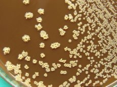
Cultural properties of Nocardia
Nocardia grow well on simple nutrient media (MPA, MPB, Sabouraud's medium, etc.). The optimum temperature for growth is 28-37 °C. On liquid media, they form a thin transparent film resembling a spreading drop of fat; they gradually acquire a creamy-yellow color. Bottom growth in the form of lumps of cotton wool or dense grains is possible. On dense media, after 45-72 hours, they form small smooth moist colonies of a doughy consistency. After 72 hours, the surface of the colonies changes; on the 10th-14th day, they take on the appearance of a raised and twisted center and scalloped edges. They produce pigments from cream to red, which diffuse into the nutrient medium. Bacteria of the 1st group form soft, pasty and mucous colonies, the 2nd - pasty or oily, the 3rd - dry leathery colonies.
Biochemical activity is quite high.
Ecological niche of Nocardia
Nocardia are widespread in soil and organic substrates. They are not representatives of the normal microflora of the human body, although they are sometimes isolated from clinically healthy people. Resistance to the environment is high.
Antimicrobial susceptibility
Nocardia are sensitive to gentamicin and chloramphenicol, commonly used antiseptics and disinfectants.
Pathogenesis of nocardiosis
Nocardia cause opportunistic infection. The pathogen is captured by alveolar macrophages, in the cytoplasm of which it remains viable, blocking the fusion of phagosomes with disosomes and inhibiting the synthesis of lysosomal enzymes. Persistence of the pathogen leads to the development of inflammation with the formation of multiple confluent abscesses and granulomas. Infection of the subcutaneous tissue develops when the pathogen enters the wound and is characterized by the development of purulent inflammation. In immunodeficient individuals, disseminated infections may develop.
Epidemiology of nocardiosis
The source of infection is soil. The mechanism of transmission is contact, the route of transmission is wound. Aerogenic transmission of the pathogen by airborne droplets or airborne dust and transmission by the alimentary route with ischium through damaged mucous membranes of the gastrointestinal tract are also possible. Susceptibility to nocardia, as to all opportunistic microbes, is low in individuals with normal immune status and increased in immunodeficient hosts.
 [ 12 ], [ 13 ], [ 14 ], [ 15 ], [ 16 ], [ 17 ], [ 18 ], [ 19 ], [ 20 ]
Symptoms of nocardiosis
Nocardioses are opportunistic human infections caused by nocardia, which are characterized by predominant damage to the lungs and subcutaneous tissue with the development of purulent-granulomatous inflammation.
They are rare infectious diseases. Every year, 1.5-2 thousand cases of the disease are registered in the world, more than half of them in people with immunodeficiencies. The main forms of damage are pulmonary and subcutaneous nocardiosis. The most common are pulmonary damage caused by Nocardia aateroides, and subcutaneous damage caused by Nocardia brasiliensis.
In case of pulmonary damage, multiple confluent abscesses and granulomas are formed in the lung parenchyma. The inflammatory process often involves the mediastinal organs, soft tissues of the chest, etc. The disease is especially dangerous for people with immunodeficiencies, who often develop disseminated infections accompanied by damage to the central nervous system, meningeal phenomena and paralysis. In disseminated forms, damage to the skin, lymph nodes, liver and kidneys is possible.
Subcutaneous tissue infections are characterized by the development of pustules at the site of pathogen penetration. As the disease progresses, abscesses and granulomas form, which resemble cutaneous actinomycosis.
Immunity has not been sufficiently studied.
Laboratory diagnostics of nocardiosis
The material for the study is sputum, pus, tissue biopsy. Microscopic and bacteriological methods are used for diagnosis. Usually, diamine is diagnosed bacterioscopically by detecting non-septate hyphae in the material being studied. The final diagnosis is established based on the isolation of the pathogen.