Medical expert of the article

New publications
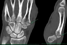
MRI of the wrist joint.
Last reviewed: 03.07.2025

All iLive content is medically reviewed or fact checked to ensure as much factual accuracy as possible.
We have strict sourcing guidelines and only link to reputable media sites, academic research institutions and, whenever possible, medically peer reviewed studies. Note that the numbers in parentheses ([1], [2], etc.) are clickable links to these studies.
If you feel that any of our content is inaccurate, out-of-date, or otherwise questionable, please select it and press Ctrl + Enter.
The wrist joint has many features: despite its small size, this joint is subject to constant and significant loads. If the wrist begins to bother and the patient consults a doctor, then an analysis of clinical symptoms alone will not be enough. It is necessary to connect additional instrumental diagnostics - in particular, the doctor may prescribe an MRI of the wrist joint. Often, only on the basis of MRI results can the doctor identify the disease.
Indications for the procedure
Medicine knows quite a lot of diseases and injuries that can disrupt the functional ability of the wrist and hand. In order to correctly diagnose and prescribe further treatment, differential diagnostics are carried out - including using such a procedure as magnetic resonance imaging.
MRI of the wrist joint is indicated for the following possible diseases:
- Developmental anomalies.
Often, anomalies in the development of joint elements are discovered by chance - especially if such defects do not cause serious functional limitations. Doctors sometimes manage to diagnose concrescence (connection) of small bone elements with each other, which to a certain extent reduces the motor amplitude in the wrist joint.
In addition, hypoplasia or aplasia of individual bones or their parts can be detected. With such an anomaly, on the contrary, pathological mobility occurs in the joint. Additional elements in the wrist are found less often.
Congenital pathologies such as dislocation and subluxation of the wrist joint also lead to impaired functionality of the hand. Fortunately, these defects are not very common and are treated surgically.
- Injuries.
Most often, traumatologists have to diagnose bruises, internal hematomas or hemarthrosis in the wrist joint. Dislocations of the joint are quite rare, since in most cases they are detected against the background of a fracture of the radius or styloid process.
Among the injuries to the bone inside the joint, the most common is a fracture of the distal epiphysis of the radius, or its fracture in a characteristic place (the so-called Colles fracture). Often, such an injury occurs against the background of damage to the head of the ulna, the styloid process and the joint disc.
- Inflammation of the joints.
Arthritis of the wrist joint can be acute or chronic, infectious or post-traumatic. Among chronic arthritis, MRI is often required for such diseases as rheumatoid and reactive arthritis, joint damage in patients with tuberculosis or brucellosis.
- Arthrosis.
After various injuries or joint inflammations, arthrosis with deformation of the wrist joint may develop. This pathology is rare, but it is extremely important to diagnose it in a timely manner. With long-term deforming arthrosis, a gradual increase in stiffness and deformation is observed, and patients complain of frequent crunching and pain during movements.
- Kienböck's disease.
Osteonecrosis of the lunate bone is also called carpal osteochondritis or osteochondropathy, lunatomalacia, avascular necrosis, or aseptic necrosis of the wrist. The essence of the disease is limited movement in the wrist joint (some patients cannot even clench their fingers into a fist). Such a pathology is not considered rare.
- Diseases of the soft tissues of the wrist joint.
Such diseases affect the soft tissues of the joint, and an MRI procedure is often prescribed for their diagnosis:
- inflammation of the joint capsule;
- tendovaginitis and tendinitis;
- periarthritis;
- ligamentitis.
It is also important to remember that tumor processes can also form in the wrist area - for example, we can talk about chondroma, osteosarcoma, osteoma, etc. Therefore, with any such suspicions, the doctor can prescribe the patient such a type of diagnostics as MRI of the wrist joint.
Preparation
In the vast majority of cases, MRI of the wrist does not require any special preparation: the joint is perfectly visualized. If contrast is used, the doctor may warn about the need to perform the procedure on an empty stomach. It is necessary to consult with the doctor in advance to identify contraindications to the procedure: during the consultation, the medical specialist will explain to the patient all aspects of the study.
First of all, the doctor should pay attention to the following questions:
- does the patient have any contraindications to this type of diagnostics (such contraindications may vary, depending on what type of MRI machine is used – closed or open);
- Is it necessary to use contrast before the procedure, and if so, is the patient allergic to the contrast agent;
- Is there a need for additional sedatives or analgesics before the procedure?
It is worth noting that MRI of the wrist joint is considered a relatively common type of diagnostics, and is often performed on open-type devices. An open procedure significantly simplifies preparation and reduces the likelihood of stress for the patient. The type of device does not affect the accuracy and informativeness of the results.
 [ 7 ]
[ 7 ]
Technique MRI of the wrist joint.
In order for the patient not to encounter any surprises during the MRI procedure of the wrist joint, he should at least have a general idea of how the examination is carried out. Thus, the standard technique for carrying out the procedure looks like this:
- The patient removes his outer clothing, as well as all foreign objects (jewelry, watches, hearing amplifiers, etc.);
- is placed horizontally on a special pull-out couch, which is then pushed into the apparatus (during an open procedure, the patient simply assumes a sitting position and places the arm being examined inside the apparatus);
- During the required time (approximately 20 minutes) the patient must remain completely still.
After the procedure is completed, the patient must remain in the office for some time so that the doctor can make sure that everything went well and there is no need for additional manipulations.
If sedation or anesthesia was additionally used before the MRI, then after the procedure one of the relatives must accompany the patient - home or to the hospital. The patient is prohibited from driving independently after sedation.
What does an MRI of the wrist show?
A high-quality image of the wrist joint in MRI images is obtained only in the most uniform magnetic field, which cannot be achieved without additional alignment. Therefore, in the MRI machine, special shimming coils are added to the basic magnet, creating gradients that compensate for technical magnetic inhomogeneity and level the impact on the patient's field. The coils create gradient pulses in three spatial directions and are coordinated by a system of amplifiers.
The radio pulse sensor (also known as the transmit coil of the MRI machine) transmits waves with a resonant frequency, modulating them into pulses of a certain type.
The receiving coil is a sensitive antenna installed counter to the direction of the base magnetic field. To avoid interference, the MRI magnet is placed in a special chamber (the so-called "cage") made of copper or aluminum sheets or rods. The received signal is converted by an analog-to-digital transformer into digital form, after which it is transmitted to the computer. The image is reconstructed and a tomogram is displayed on the monitor.
The described principle of operation of the MRI apparatus helps to give an accurate assessment of the condition of soft tissues, cartilage, and ligaments. MRI of the wrist joint is less informative in case of bone tissue damage.
What does an MRI of the hand and wrist joint provide?
- During the examination, it becomes possible to obtain a detailed image of the problem area. Therefore, MRI of the wrist joint is especially effective for early diagnosis of tumor and inflammatory processes.
- MRI helps to examine areas that are not visualized using CT - for example, when the required area is covered by bone tissue, or due to the poor sensitivity of CT to altered tissue density.
- MRI allows us to evaluate not only the structure of tissues, but also the quality of their functioning (for example, we can record the speed of blood flow).
The results obtained during the MRI of the wrist joint are given to the patient or passed on to the doctor. This can happen in a couple of hours or the next day after the procedure.
Contraindications to the procedure
Restrictions to performing MRI of the wrist joint can be absolute and relative (i.e., temporary). Absolute restrictions are:
- the presence of a foreign metal object in the body;
- the presence of a metal or electromagnetic implant or prosthesis;
- the presence of a pacemaker, insulin pump.
If an MRI of the wrist with contrast is required, such a procedure cannot be performed on patients with renal insufficiency or hypersensitivity to the ingredients of the contrast agent.
Relative restrictions may include:
- pregnancy during the first trimester;
- mental disorders, panic attacks, schizophrenia, fear of enclosed spaces (when using a closed-type device);
- severe decompensated conditions;
- the presence of tattoos with metal-containing dyes;
- severe pain, itching - that is, symptoms that prevent the patient from remaining motionless for a long time;
- a state of alcohol or drug intoxication.
Obesity is also considered a contraindication for closed-type devices, since the MRI camera has limitations on the patient's body weight and volume. It is believed that the maximum patient weight for the procedure should not exceed 150 kg. Open-type devices do not have such a limitation.
Childhood cannot be a contraindication as such. However, it is sometimes difficult to diagnose children - primarily because children simply cannot remain motionless for a long time. If there is an urgent need to conduct an MRI of the wrist joint in a child, then preliminary use of sedatives or anesthesia is possible.
Complications after the procedure
MRI of the wrist joint - with contrast or in the usual way, is an important diagnostic study, and it is very important for the patient to understand how exactly it affects the body. It should be noted that to date, there is not a single proven fact of the harmful effect of the procedure on health. Most experts agree that MRI does not affect the body's functionality in any way - either in one direction or another.
Some people without proper experience may claim that MRI (including wrist) can harm health due to prolonged exposure to a powerful magnetic field. In fact, this is not true: the magnet interacts only with hydrogen atoms, and with nothing else. It turns out that the water molecules present in the body simply line up in parallel with the magnetic field, which cannot affect the condition and functions of the body in any way.
The principle of image formation can also be explained. When exposed to a magnetic wave, previously aligned atoms begin to vibrate, emitting energy, which is then converted into an image. Thus, both the magnetic field and the radiation are completely safe. Experts are confident that the MRI procedure of the wrist joint can be repeated several times if necessary: this will not affect the health of the body in any way.
Does the type of MRI machine used matter for patient safety? Are there any health complications that can arise from an open or closed procedure?
The closed device looks like a special cylindrical chamber, open on both sides. The patient "enters" this chamber, lying on a mobile couch, and stays there for a certain amount of time. If a person suffers from claustrophobia, then do not tempt fate: it is better to refuse a closed procedure in favor of an open-type device. Otherwise, some problems of a corresponding nature may indeed arise.
Both open and closed devices can cause complications if the patient does not warn the doctor about the presence of metal implants in his body, about the presence of an allergy to the contrast agent, as well as about other possible contraindications to the study.
- If the patient does not warn the doctor about the presence of a severe kidney disease, then under the influence of the magnetic field and contrast agent, the pathology can transform into nephrogenic fibrosis.
- If the patient does not remove metal objects from the body before the procedure, some skin problems may arise in the form of superficial tissue damage.
- If the patient has a device such as a pacemaker, it may simply stop working during the procedure: the outcome of such a situation is not difficult to predict.
- If the patient has an allergic predisposition to the components of the contrast agent, then after the administration of the latter, the following may be observed:
- difficulty breathing;
- increased heart rate;
- skin rash, swelling, etc.
It is highly advisable to do an allergy test before the contrasting procedure. This will help prevent very unpleasant consequences and complications.
Care after the procedure
Almost all patients can go home on their own after an MRI of the wrist: no special regimen or care is required for them. The doctor may ask the patient to wait for about an hour in the next room to receive the images and a medical report. In some cases, the results are transmitted directly to the attending physician.
Some clinics practice recording MRI data on a USB storage device. In such a case, the patient waits less time.
If the patient was given sedatives or anesthesia before the MRI, then a close person should accompany him/her after the procedure. Independent movement of the patient after sedation or anesthesia - on foot or in transport - is prohibited. If the patient's health worsens, you should immediately contact a doctor.
[ 13 ]
Reviews
MRI is considered one of the most modern, accurate and safe diagnostic procedures. Such diagnostics are informative, painless and can be used even in pediatrics. In the field of proven safety, MRI is a more preferable method of examination, in contrast to X-ray or computed tomography. Moreover, X-ray does not have such accuracy: this method is more suitable for diagnosing injuries of the skeletal system. Computed tomography is more informative, but also involves the use of X-ray irradiation. Therefore, in this aspect, the use of magnetic resonance imaging is preferable.
The relatively high cost is perhaps the only obvious "minus" of MRI of the wrist joint. However, most patients still choose this type of diagnostics because of its informativeness and safety.