
Medical expert of the article

New publications
MRI of soft tissues: what the test shows, when it is prescribed, and whether contrast is needed
Last updated: 11.04.2026

All iLive content is medically reviewed or fact checked to ensure as much factual accuracy as possible.
We have strict sourcing guidelines and only link to reputable media sites, academic research institutions and, whenever possible, medically peer reviewed studies. Note that the numbers in parentheses ([1], [2], etc.) are clickable links to these studies.
If you feel that any of our content is inaccurate, out-of-date, or otherwise questionable, please select it and press Ctrl + Enter.
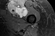
Soft tissue magnetic resonance imaging (MRI) is an imaging technique that uses a powerful magnetic field, radiofrequency pulses, and computer signal processing to produce detailed images of internal structures without ionizing radiation. This technique is particularly valuable for soft tissue structures because it often more accurately reveals the differences between normal and abnormal tissue than other methods, and is also better able to detect areas that may be partially obscured by bone in other imaging studies. [1]
In clinical practice, the term "soft tissue magnetic resonance imaging" covers a variety of examinations: those of the neck, extremities, pelvis, abdominal wall, gluteal region, feet, hands, and other areas. The specific protocol always depends on the location of the problem and the specific issue being addressed: ruling out a tumor, clarifying inflammation, assessing trauma, the relationship of the mass to vascular and neurological structures, or monitoring treatment. [2]
The most important thing for patients to understand in advance is that soft tissue magnetic resonance imaging (MRI) is very informative, but it is not a universal first step for every lump, pain, or swelling. For small, superficial lesions, an ultrasound is often the first test, and MRI is used when more anatomical detail and a more precise local assessment are needed. [3]
What is soft tissue magnetic resonance imaging and what does it show?
Soft tissue is not a single organ, but a large group of structures. In practical terms, this includes muscles, subcutaneous fat, fascia, tendons, ligaments, neurovascular bundles, lymph nodes, soft tissue structures, and many regional structures that are not dense bone tissue. This is why the same examination can be used for a suspected hip tumor, an unclear inflammatory process in the leg, and for assessing a deep soft tissue mass in the shoulder. [4]
The strength of this method lies in its high soft tissue contrast. The radiologist obtains images in multiple sequences and planes, allowing them to see not only the presence of a lesion, but also its shape, size, depth, internal structure, boundaries, and relationship to fascia, muscles, vessels, nerves, and bone. For many soft tissue lesions, this is key information, which subsequently determines the choice of treatment strategy. [5]
In oncology and the diagnosis of soft tissue tumors, magnetic resonance imaging is particularly important because it is considered the method of choice for local characterization and staging of most soft tissue tumors. European guidelines for soft tissue tumor imaging emphasize that this method is best suited for local assessment in most cases, while computed tomography is used in specialized situations. [6]
However, this method has reasonable limitations. Magnetic resonance imaging can very well describe a lesion, but it is not always able to distinguish benign from malignant processes with 100% accuracy from an image. Therefore, if suspicious signs are present, a biopsy is often the next step, and recommendations emphasize that high-quality magnetic resonance imaging should be performed before a biopsy, not after, if it can improve the characterization of the lesion. [7]
Another important point is that this method is not equally useful for every problem. It's excellent for assessing soft tissue, but it's not always the best start for a small, superficial lump, isn't always necessary without a clinical question, and doesn't replace a doctor's examination. A properly ordered examination is more valuable than simply "getting an MRI just in case." [8]
| What does soft tissue magnetic resonance imaging evaluate? | Why is this important? |
|---|---|
| Size and shape of the hearth | Helps to understand the scale of the process |
| Depth and connection with fascia | Affects suspicion of aggressiveness |
| Internal structure | Allows you to see fluid, fat, necrosis, hemorrhage |
| Connection with muscles, blood vessels, nerves | Needed for treatment planning |
| The presence of concomitant edema and inflammation | Helps to differentiate variants of the pathological process |
The table is compiled based on data from the ACR, RadiologyInfo and European guidelines for soft tissue tumors. [9]
When is soft tissue magnetic resonance imaging really necessary?
If the lesion is superficial, particularly small and accessible to the transducer, the first steps are often ultrasound and radiography. The American College of Radiology indicates that for a superficial mass, ultrasound and radiography are usually the initial appropriate approach, rather than magnetic resonance imaging. This is important because patients often expect the "most powerful" examination right away, even though the initial procedure is simpler. [10]
If the lesion is deep, located in a difficult-to-reach area, poorly characterized by ultrasound, or initial studies have not yielded a clear answer, the situation changes. In this case, after non-diagnostic radiography and ultrasound, magnetic resonance imaging (MRI) with and without contrast is usually considered an appropriate next step, and a non-contrast examination may be an acceptable alternative in certain cases. [11]
Particular attention is required if the soft tissue lesion is enlarged, deep-seated, painful, or larger than 5 cm. National and sarcoma referral pathways consider such signs to be warning signs. This does not necessarily mean that every such lump is malignant, but it does mean that further imaging and consultation should not be delayed. [12]
If a soft tissue tumor is suspected, this method is valuable not only for the diagnosis itself but also for planning further steps. European guidelines emphasize that ultrasound remains a good initial triage method for accessible and small tumors, but if the diagnosis is unclear and a tumor is suspected, magnetic resonance imaging should precede biopsy if it adds information to characterize the lesion. [13]
After treatment of soft tissue sarcomas, magnetic resonance imaging also plays a major role in local surveillance. Updated guidelines from the European Society of Musculoskeletal Radiology explicitly state that this method is generally considered the method of choice for locoregional surveillance of soft tissue sarcomas, while chest computed tomography is used to monitor metastases. [14]
| Clinical situation | Usually the preferred first step | When is magnetic resonance imaging needed? |
|---|---|---|
| A small superficial lump | Ultrasound examination, radiography | If the answer is unclear |
| Deep education | X-ray, sometimes ultrasound | Often required for clarification |
| Unclear initial research result | Clinical re-evaluation | Usually the next logical step is |
| Suspicion of sarcoma | Urgent visualization along the route | Before biopsy for better local mapping |
| Follow-up after treatment of soft tissue sarcoma | Planned observation | Often the method of choice for a local area |
The table is based on the ACR criteria, RadiologyInfo and European guidelines for soft tissue tumors. [15]
How the study is conducted and how to prepare for it
Preparation begins not with food, but with properly informing the staff. Before the examination, you must disclose any previous surgeries, implants, metal in your body, allergies, pregnancy, as well as any claustrophobia or anxiety. RadiologyInfo specifically emphasizes that most orthopedic implants are safe, but any device or metal should be disclosed in advance. [16]
Before entering the scanner area, you must remove jewelry, watches, hearing aids, and removable metal objects, and you may sometimes be asked to change into a hospital gown. The reason is simple: the strong magnetic field can affect metal objects, and some devices may heat up, shift, or distort the image. For this reason, MRI safety is based on strict questionnaires and screening before the examination, not during it. [17]
The scan itself typically lasts approximately 30-60 minutes, although the time depends on the area of the body and whether contrast is needed. It is important to remain still during the scan, as movement degrades image quality. For some torso scans, short breath holds of 15-25 seconds may be required after contrast. [18]
The procedure is generally tolerable for most patients. The examination is generally painless, but the machine is noisy, and some people experience a feeling of confinement. RadiologyInfo notes that some patients feel constrained in the machine tunnel, and for severe claustrophobia, a mild sedative is sometimes prescribed beforehand. [19]
After the examination is complete, a radiologist evaluates the images and issues a report. In many clinics, the results are sent to the patient's personal account, but the final interpretation should be discussed with the attending physician, who is familiar with the clinical context. Sometimes, after the initial report, a repeat examination, additional sequence, or follow-up is required, and this is not a sign of error, but rather part of the normal diagnostic logic. [20]
| Preparing for the study | What is usually required |
|---|---|
| Report implants and metal | Necessarily |
| Remove jewelry and electronics | Necessarily |
| Change into hospital gown | Often yes |
| Get a kidney function test | Sometimes, if contrast is planned |
| Discuss claustrophobia in advance | Desirable |
| Arrange for support after a sedative | If the drug is prescribed |
The table is compiled from data from RadiologyInfo and materials on the safety of magnetic resonance imaging. [21]
When is contrast needed and how safe is it?
Contrast is not always necessary for soft tissue magnetic resonance imaging (MRI). However, when assessing soft tissue lesions, it often helps better understand the structure of the lesion, its blood supply, the presence of septa, necrotic zones, inflammatory changes, and its relationship to surrounding tissue. In the ACR criteria, for situations where initial radiography and ultrasound are inconclusive, MRI with and without contrast is generally considered the appropriate next test. [22]
Contrast agents used in magnetic resonance imaging (MRI) are most often gadolinium-based. According to RadiologyInfo, allergic reactions to these agents are less common than to iodine-containing contrast agents used in CT scans. However, absolute safety is not guaranteed, so a patient's allergy history and underlying medical conditions are always reviewed before administration. [23]
The most important risk group is patients with severe renal impairment. RadiologyInfo notes that nephrogenic systemic fibrosis is a recognized complication of gadolinium-containing agents, but with newer agents it is extremely rare and usually associated with serious renal pathology. The ACR also recommends using gadolinium with extreme caution in patients with high renal risk and, if contrast is essential, choosing safer agents at the lowest dose sufficient for diagnosis. [24]
Pregnancy requires a special approach. RadiologyInfo reports that magnetic resonance imaging has been used since the 1980s, and there have been no reports of harm to the pregnant woman or fetus from the method itself. However, in the first trimester, the examination is only performed when the benefits outweigh the potential risks. Gadolinium contrast during pregnancy is only permitted in critical cases where it is essential to obtain information that impacts pregnancy management or treatment. [25]
When breastfeeding, restrictions are more relaxed. The ACR states that less than 0.04% of the maternal gadolinium dose is excreted into breast milk, and less than 0.0004% of the maternal intravascular dose is absorbed systemically by the infant, so continuing breastfeeding after such contrast is generally safe. Formally, the decision can be discussed on an individual basis, but routine discontinuation of breastfeeding is not required. [26]
| Question about contrast | Practical answer |
|---|---|
| Does everyone need contrast? | No |
| How often is it useful for soft tissue lesions? | Yes |
| Allergy to gadolinium is possible | Yes, but less frequently than with iodine-containing contrast |
| When caution is especially important | In severe renal failure and pregnancy |
| Should I stop breastfeeding after gadolinium injection? | Usually no |
The table is based on data from the ACR Manual on Contrast Media and RadiologyInfo. [27]
How does magnetic resonance imaging of soft tissue differ from ultrasound and computed tomography?
Ultrasound, computed tomography, and magnetic resonance imaging are not directly competitive in all situations. Ultrasound is convenient as a rapid, accessible, and dynamic primary method for small superficial lesions. European guidelines for soft tissue tumors explicitly state that for accessible and small suspected soft tissue tumors, ultrasound remains the best primary triage method. [28]
Magnetic resonance imaging (MRI) is particularly powerful when it comes to understanding the depth of a tumor, its internal structure, and its spread through soft tissue. European guidelines state this very clearly: MRI is the method of choice for characterization and local staging of most soft tissue tumors. When it comes to the precise anatomy of the lesion, including its fascia, muscles, nerves, vascular proximity, and relationship to bone, MRI typically provides more information. [29]
Computed tomography remains important, but in different roles. It is useful when magnetic resonance imaging is contraindicated, when additional calcifications or bone changes are needed, or when the patient is unable to undergo magnetic resonance imaging. The ACR criteria specifically state that when magnetic resonance imaging is contraindicated, contrast-enhanced computed tomography is usually the next appropriate test. [30]
There are also technical limitations to the method itself. High image quality depends on the ability to lie still, occasionally perform short breath holds, and compatibility with implants must be confirmed in advance. RadiologyInfo also notes that in some patients, a large body size, severe pain, anxiety, or an inability to lie still reduce the diagnostic value of the study. [31]
Therefore, the correct choice of method is always based not on the question "what's best in general," but on the question "what's best for this specific area and this specific problem." For a small, superficial lipoma, an ultrasound may be sufficient. For a deep, unclear mass in the thigh or shoulder, magnetic resonance imaging is usually the preferred method. If magnetic fields are contraindicated, computed tomography (CT) is a reasonable alternative. [32]
| Method | Strengths | Weaknesses |
|---|---|---|
| Ultrasound examination | Fast, affordable, good for small superficial lesions | Limited in deep and complex formations |
| Magnetic resonance imaging | Best soft tissue contrast, precise local anatomy, no ionizing radiation | Longer, more expensive, sensitive to movement and safety restrictions |
| Computed tomography | Useful for contraindications to magnetic fields, clearly sees bone and calcifications | Uses ionizing radiation, soft tissue contrast is usually lower |
The table is compiled based on data from RadiologyInfo, ACR and European guidelines for soft tissue tumors. [33]
What does the conclusion mean and when should you not delay further examination?
In the report, the radiologist typically describes the location, size, depth, and signal from the lesion on different sequences, the presence of contrast enhancement, and its relationship to fascia, muscles, vessels, nerves, and bone, and then formulates the most probable diagnostic sequence. Modern materials describing soft tissue tumors emphasize that these parameters are critical for further evaluation and treatment. However, the final verdict is not always definitive, especially when dealing with an ambiguous mass. [34]
It's important for patients to remember that a good conclusion doesn't always end with the words "benign" or "malignant." Often, the wording is more cautious: for example, "data suggest a lipomatous lesion without aggressive features" or "a soft tissue tumor cannot be ruled out; consultation with a specialized center is recommended." This restraint is not a flaw in the method, but rather reflects sound clinical practice. [35]
Particularly careful attention should be paid to formations that are growing, located deep, painful, or larger than 5 cm. These are the signs considered alarming in sarcoma lesions. They do not automatically indicate cancer, but they do indicate that the examination should be carried out correctly and without delay. [36]
If the report indicates the need for a follow-up examination, dynamic monitoring, or biopsy, this should not be delayed. European guidelines specifically emphasize that suspicious or likely malignant tumors should be biopsied, while large and indeterminate lesions are best discussed with a reference tumor center prior to intervention. Magnetic resonance imaging in this context provides a map of the process, but the final approach is determined by the overall clinical picture. [37]
Urgency is especially important if, in addition to the mass itself, pain, limited movement, numbness, rapid growth, nocturnal symptoms, or concomitant weight loss occur. In layman's terms, this is the primary guideline: not every lump is dangerous, but a rapidly growing, deep, and painful mass should never be left unassessed. [38]
| Wording in the conclusion | What does this usually mean? |
|---|---|
| Signs of a benign tumor | Monitoring or scheduled treatment is often required. |
| Ambiguous soft tissue mass | Consultation and further examination are required. |
| Suspected aggressive formation | A specialized route is needed |
| A biopsy is recommended | Visualization is not sufficient for a definitive diagnosis. |
| Dynamic control is recommended | It is important to compare changes over time |
The table is based on materials describing soft tissue tumors and clinical guidelines for their routing. [39]
FAQ
Are soft tissue magnetic resonance imaging and magnetic resonance imaging with contrast the same thing?
No. The basic examination can be performed without contrast, and contrast is added when it is necessary to better evaluate the structure of the lesion, its vascularity, the area of inflammation, or a possible tumor. After unclear initial studies for a soft tissue mass, the ACR often considers both a non-contrast and a contrast-enhanced examination most appropriate. [40]
Can an MRI scan tell for sure if it's cancer?
Not always. The method very well describes the formation and its prevalence, but in some cases it is impossible to definitively distinguish a benign process from a malignant one based on the image, so if there are suspicious signs, a biopsy is required. [41]
If there is metal in the body, is the examination prohibited?
Not necessarily. Many orthopedic implants pose no problems, but some devices and individual metal objects can be dangerous or significantly impair image quality. This is why a thorough interview and confirmation of the device's compatibility with MRI are essential before the examination. [42]
Is it possible to do the test during pregnancy?
Sometimes yes, if the benefit to the mother or fetus outweighs the potential risks, but in the first trimester, its use is approached with particular caution. Gadolinium contrast is permitted during pregnancy only when critical information cannot be obtained without it. [43]
Should I stop breastfeeding after gadolinium?
Usually not. The ACR points out that the amount of gadolinium that enters breast milk and is then actually absorbed by the baby is very small, so it is usually safe to continue breastfeeding after the test.[44]
What to do if you have severe claustrophobia?
This should be discussed in advance. For some patients, an open device is suitable, while for others, a mild sedative is needed before the examination. The main thing is to not hide the problem, as a calm, motionless position directly affects the quality of the images. [45]
Which is better for a soft tissue lump: ultrasound or magnetic resonance imaging?
It depends on the situation. For a small, superficial, and accessible lump, ultrasound is often the appropriate first step. For a deep, unclear, or suspicious mass, magnetic resonance imaging is usually needed. [46]
Key points from experts
Iris-Melanie Noebauer-Huhmann, MD, Professor, Head of Clinical Research in the Division of Neuroradiology and Musculoskeletal Radiology at the Medical University of Vienna. As the scientific guarantor of the European guidelines for soft tissue tumors in adults, she presents a very practical position: ultrasound remains a good initial triage method for small, accessible lesions, but magnetic resonance imaging is the method of choice for characterization and local staging of most soft tissue tumors, and is best performed before biopsy. [47]
Hillary W. Garner, MD, professor of radiology at Mayo Clinic and an expert in musculoskeletal imaging, reflects a key practical principle for routine workup: if radiography and ultrasound are inconclusive, magnetic resonance imaging (MRI) with or without contrast is usually the appropriate next step. [48]
Ivan Pedrosa, MD, PhD, vice chair for strategy and innovation and professor in the Department of Radiology at MD Anderson Cancer Center, is the author of the updated ACR magnetic resonance imaging safety guideline. His key message is that modern magnetic resonance imaging is safe only when screening protocols are strictly followed, especially in patients with implants, devices, and potential risk factors associated with contrast. [49]
Daniel E. Wessell, MD, PhD, is a radiologist and vice chair of scientific affairs and director of the Mayo Clinic Musculoskeletal Radiology Fellowship. His expertise in tumors, infection, trauma, and advanced magnetic resonance imaging (MRI) and computed tomography (CT) reflects the modern idea that the value of a study is determined not only by the availability of images but also by the quality of the protocol, the completeness of the description, and the appropriate patient referral. [50]
Conclusion
Magnetic resonance imaging of soft tissues is one of the most informative methods for assessing soft tissue structures, especially when it is necessary to clearly visualize the depth of a process, its internal structure, and its relationship to surrounding tissues. It does not use ionizing radiation, provides excellent visualization of soft tissues, and is often the method of choice for clarifying unclear or suspicious soft tissue lesions. [51]
However, this is not a universal first step for every complaint. For small superficial lesions, ultrasound is often the first step, and if magnetic field examination is contraindicated, CT scanning is the next step. The examination is most beneficial when prescribed for the correct indications, performed with the correct protocol, and interpreted in the clinical context, not in isolation from the patient's symptoms. [52]