Medical expert of the article

New publications
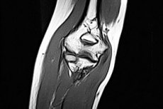
MRI of the elbow
Last reviewed: 03.07.2025

All iLive content is medically reviewed or fact checked to ensure as much factual accuracy as possible.
We have strict sourcing guidelines and only link to reputable media sites, academic research institutions and, whenever possible, medically peer reviewed studies. Note that the numbers in parentheses ([1], [2], etc.) are clickable links to these studies.
If you feel that any of our content is inaccurate, out-of-date, or otherwise questionable, please select it and press Ctrl + Enter.
 [
[ Indications for the procedure
Indications for magnetic resonance imaging include injuries, as well as inflammatory and degenerative diseases of this joint, accompanied by pain and limited mobility.
It is difficult to overestimate the role of high-contrast images and detailed visualization of the structures of the elbow joint using MRI, because it has a complex structure - three simple joints (block-shaped humero-ulnar, ball-and-socket humero-radial and cylindrical radio-ulnar), which articulate three bones, located in a common joint capsule.
Since it is the MRI of the elbow joint that provides the most complete information regarding its condition, this method becomes the main tool for differential diagnostics. In addition, such examination is necessary before surgical intervention - osteosynthesis, arthroplasty or endoprosthetics, as well as for assessing their results.
What an MRI of the elbow joint shows – the articular surfaces of the bones that form the joint and the cartilage covering them, the condyles and epicondyles, the joint capsule and its synovial membrane, ligaments, tendons, soft tissues surrounding the joint, blood vessels and nerves – minimizes the risk of an erroneous diagnosis.
Thanks to the layer-by-layer three-dimensional reconstruction of the image of all internal structures of the joint, the MRI anatomy of the elbow joint of a specific patient is determined. The specialist compares it with the images in electronic atlases of normal MRI sections and identifies deviations.
Based on the analysis of these deviations, the following is diagnosed:
- fractures of the olecranon, coronoid process of the ulna, neck and head of the radius with rotational displacement, crushing, fragments, ligament rupture;
- inflammation of the elbow joint (arthritis), its joint capsule (bursitis) or synovial membrane (tenosynovitis);
- dystrophy of articular cartilage and development of arthrosis (osteoarthrosis);
- inflammation of the periosteum, tendons and ligaments in the area of the epicondyles of the humerus - epicondylitis of the elbow joint;
- tunnel syndrome of the elbow joint (cubital tunnel syndrome).
MRI reveals the presence of post-traumatic, periosteal or degenerative-dystrophic osteophytes (bone growths) in the joint and surrounding tissues.
Technique MRI of the elbow
If the examination is carried out on a closed tunnel-type tomograph, the patient lies on his back (or stomach), stretching out his arm. With an open tomograph (without a tunnel chamber), the examination is carried out in a sitting position.
But in any case, the positioning of the limb during MRI of the elbow joint is carried out according to the guidelines for scanning parameters and patient placement techniques - with fixation of the head and limbs to ensure complete immobility.
An MRI scan of one elbow joint can take 15 to 25 minutes.
Contraindications to the procedure
It is contraindicated to perform an MRI examination of the elbow joint (and MRI scanning in general) if the patient has an implanted artificial pacemaker, insulin pump, or cochlear implants, metal dental crowns and prostheses, vascular stents and clips, intraosseous pins, etc.
MRI is not performed in cases of claustrophobia or during the first trimester of pregnancy.
For tunnel-type tomographs, the patient's body weight is limited to 120-130 kg.
Complications after the procedure
In some cases - with higher EMF voltage of powerful tomographs or violations of the examination protocol regarding its duration - complications after the procedure are possible in the form of quickly passing dizziness, the appearance of a metallic taste in the mouth or involuntary twitching of individual muscles.
As the majority of patients testify, MRI of the joints did not affect their well-being in any way and made it possible to identify the exact cause of problems with the elbow joint.