Medical expert of the article

New publications
Liver fluke
Last reviewed: 04.07.2025

All iLive content is medically reviewed or fact checked to ensure as much factual accuracy as possible.
We have strict sourcing guidelines and only link to reputable media sites, academic research institutions and, whenever possible, medically peer reviewed studies. Note that the numbers in parentheses ([1], [2], etc.) are clickable links to these studies.
If you feel that any of our content is inaccurate, out-of-date, or otherwise questionable, please select it and press Ctrl + Enter.
The liver fluke is a parasite that affects humans and can cause non-specific symptoms that are difficult to explain and treat. This disease is more common than it is diagnosed, so it is important to have an idea of the main symptoms and clinical manifestations of this pathology. Rare diagnostics also concerns other human helminthiases due to the non-specific picture and course of the disease.
All human infections caused by worms are called helminthiasis and are classified depending on the type of pathogen and the structure characteristic of this group. They are distinguished:
- Trematodes are the so-called sysuns or flatworms - the causative agents of schistosomiasis, dicrocoeliosis, opisthorchiasis, fascioliasis;
- Nematodes are roundworms that cause the following diseases: ascariasis, trichuriasis, trichinosis, enterobiasis;
- Cestodoses are tapeworms that cause taeniasis, taeniarhynchosis, cysticercosis, and hymenolipedosis.
All these worms have different transmission mechanisms, different life cycles and different preventive measures. One of the rare helminthiasis is fascioliasis, the causative agent of which is the liver fluke.
Structure of the liver fluke
The liver fluke or Fasciola hepatica has a complex life cycle and, given these features and its characteristic structure, it is classified as a flatworm.
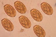
The structure of the liver fluke is quite typical for its class. The body has a lanceolate or leaf-shaped form, slightly flattened in the upper-lower direction with a characteristic dark brown color with a gray tint. At the beginning and at the end, the body narrows, has a pointed edge. The length of the parasite is no more than four centimeters, and usually about two to three and a half. Fasciola has two suckers, which are located on the front edge in the form of an oral and abdominal one, and between them is the oral opening. It is because of the presence of these suckers that these flatworms are called sysuns.
The digestive system of the liver fluke consists of two tubes that do not have an open exit, but have a blind end without an anus. This is one of the advantages that helps the helminth parasitize in the liver tract.
The hematopoietic and respiratory systems are not fully developed, which is why the liver fluke has such a characteristic localization when it enters the main and intermediate host.
The nervous system of the helminth is a nerve ring located around the pharynx and nerve fibers in the form of long strands that run along the entire body and have branches in different ends of the fluke. Such features of the nervous system allow it to respond to changes in shape and other types of irritation of the fluke's body, despite the primitiveness of such a structure.
The reproduction of the liver fluke is a rather complex process, which occurs with the change of various forms, hosts and brings a huge number of offspring from one egg. Fasciola reproduction occurs sexually and hermaphroditically. This contributes to the fact that the life cycle of the liver fluke has three generations, each with its own larvae.
Such features of the external structure and characteristic differences of the internal structure of this helminth contribute to the fact that the parasite mainly affects the liver and bile ducts, which allows it to be there without the negative influence of aggressive factors of bile, liver enzymes and cellular lysosomal enzymes. Such activity of fasciola allows it to parasitize in its main place of residence for a long time - about five years without the action of any immune factors. This is often observed in helminthiases, that their primitive structure and rather complex life cycle allows them to persist in the focus for years without pronounced clinical manifestations and a reaction from the immune system. The hosts of the liver fluke are small and large cattle, and humans are not so common.
Life cycle of liver fluke
The complexity of the structure and the various forms of reproduction of the helminth require a complex life cycle. Fasciola has three generations:
- marites with a larva called circaria;
- sporocysts with miracidium larva;
- redia with a direct path of development.
The entire development cycle begins with a hermaphroditic individual - marita. This marita lives in the host and is a sexually mature individual. It has a characteristic long body, even longer than a mature fasciola - about five centimeters. The structure of the marita makes it easy to distinguish this form from others: at the end of a long lanceolate body is the uterus with an ovary, and along the entire length of the body are ovaries. This structure contributes to the fact that it fertilizes itself due to the presence of different genetic materials of two opposite individuals. Then, after some time, fertilized eggs come out of the uterus - they have a characteristic appearance: oval, brown with a yellow tint, have a lid on one of the edges or poles of the egg. After the egg matures, larvae emerge from them - miracidia, which also have characteristic features: the entire cell is covered with outgrowths in the form of cilia, which allows it to actively swim in the water, because immediately after maturation, the miracidia needs to find a host. At the front end of the cell there is an organ that reacts to light, nerve fibers and a special substance that, upon reaching the host's body, helps dissolve its cells and penetrate inside. At the end of the cell there are special germ cells that give rise to the development of the next stage. These cells do not require fertilization, and development occurs asexually. The first host of the liver fluke is a mollusk. Miracidium penetrates the host's body with the help of this proteolytic enzyme at the front end of the cell, then migrates to the liver, where it multiplies asexually and a new phase of development is formed - a sporocyst.
The sporocyst has no characteristic structural features - it is a shapeless formation that does not have digestive, respiratory, or excretory organs. A new generation of larvae develops in them, also from ready-made germ cells that remained from the previous cycle - redia. When redia exit, the sporocyst shell ruptures and it dies, giving life to new forms. This generation already has organs - the digestive system is represented by the mouth, pharynx, and there are also organs that ensure the exit of new larvae. After one or two months, redia mature and by simple division form offspring - circaria.
Circaria are the last stage in the life cycle of the liver fluke. They already fully resemble the adult in appearance and structure. At the front end of the circaria, there are suckers, a digestive tube and nerve ganglia, organs for excreting metabolic products. A distinctive feature in the structure is the presence of a long tail in the circaria, which allows this form to leave the intermediate host and exist independently. Circaria floats freely in the water, and for further development, it attaches to plants near the shore, while covering itself with a capsule. Then a form is formed that is resistant to environmental factors and can withstand various temperature changes, drying due to the thick capsule. This form is a cyst and also has a specific name - adolescaria. The final host is animals that feed on grass or aquatic plants - these are cows, horses, goats, pigs, sheep. Getting into the gastrointestinal tract of these animals with food, the liver fluke, being at the cyst stage, dissolves its shell under the action of enzymes of the stomach and intestines, and then penetrates the wall. From the intestine, the helminth migrates through the blood of the portal vein to the liver, where it is its habitat. Coming out of the cyst, the larvae in the liver make long passages and reach the bile ducts, where their final maturation and sexual maturity occur after three months. Sometimes the helminth affects the pancreas. Then non-specific clinical symptoms begin to appear, depending on the degree of damage to the liver cells and blockage of the bile ducts.
The routes of human infection with liver fluke are limited to water bodies where intermediate hosts live – mollusks. Human infection occurs rarely, mostly by accident or in underdeveloped countries when drinking water from open water bodies infected with cysts. It can also occur when eating raw unwashed vegetables or seafood. In the human body, liver fluke is localized in the same places as in animals and causes the same changes as in the liver of mammals.
Symptoms of fascioliasis
The development of any helminthic infection is accompanied by a number of non-specific symptoms, but at the same time, sometimes characteristic symptoms are observed, which is associated with the peculiarities of the pathogenesis of the disease.
The liver fluke's defeat is characterized by its predominant localization in the liver, namely in the bile ducts, but it can also be frequently localized in the pancreas. At the larval stage, the helminth enters the liver, where it forms its liver ducts, and at the same time, the hepatocytes are destroyed and characteristic clinical manifestations are observed. Cysts, due to their thick wall, are very resistant to the action of aggressive bile secretion. When the fasciola reaches the liver ducts, it multiplies there, numerous eggs are formed, from which adult individuals later develop and destroy the duct wall, expand it and disrupt the outflow of bile and the architectonics of the liver beams. When the eggs are released with part of the bile into the gastrointestinal tract, only then can they be found in feces.
The incubation period is from one to six to eight weeks. This is the period from the time the cysts enter the human gastrointestinal tract until they migrate to the liver and begin to show clinical manifestations. When the cysts end up in the liver, they begin to multiply rapidly and mature individuals infect the cells. This period lasts until all adult helminths have dispersed throughout the liver. This period from the onset of the first symptoms until the clinical picture becomes less severe is called the acute stage.
The acute stage of liver fluke damage is characterized by non-specific manifestations such as fatigue, nausea, vomiting, headache, and an increase in temperature to both subfebrile and febrile numbers. Severe pain or just heaviness in the right hypochondrium or in the epigastrium may be disturbing, since the left lobe of the liver is most often affected. Specific signs of liver damage are the appearance of jaundice, which has a greenish tint with intense itching. Such jaundice appears due to a violation of the outflow of bile and the release of indirect bilirubin into the blood, as well as an increase in the amount of bile acids that cannot enter the intestines and have an irritating effect on the skin, that is, itching appears.
In the acute stage, symptoms of allergic manifestations are often observed, which can have varying degrees of severity from skin lesions in the form of urticaria to serious manifestations in the form of Quincke's edema. Such allergic manifestations are explained by the release of helminth metabolic products into the blood, which has a strong allergenic effect.
But such pronounced clinical symptoms do not occur in everyone and not so often. Quite often, helminthiases have an unexpressed acute stage, which makes diagnostics very difficult. Symptoms may not be so pronounced, jaundice may not appear, and the only clinical manifestations may be nausea, vomiting, pain in the hypochondrium, which is often assessed as cholecystitis, gallstone disease or simple poisoning.
After some time, on average from two to three weeks, the symptoms gradually fade and the next stage develops - chronic. The course of this stage may differ, since there are different types of damage. With continued parasitism of the helminth, a picture of chronic cholecystitis with periodic exacerbations develops. Helminths can cause a violation of the rheological state of bile and this contributes to the formation of stones and the development of cholelithiasis. Very often, small cysts of the liver fluke are not visualized on ultrasound, which does not give reason to suspect anything else. When infection of the bile ducts occurs, a picture of acute cholecystitis or cholangitis develops. All these pathologies develop against the background of only one small helminth, which can live for ten years, and treatment will be ineffective due to its non-specificity.
Diagnosis of human liver fluke infection
Due to the fact that the liver fluke has a number of non-specific symptoms, and the severity of these symptoms may be insignificant, the issue of timely diagnosis is very difficult. Often, a diagnosis indicating the location of the lesion is not made during a person's life, since the eggs are not excreted constantly and may not be in all portions of feces, and only specific diagnostics can confirm the diagnosis.
As for the anamnesis, it is very important to find out from the patient when the first symptoms appeared. It is necessary to know about possible episodes of infection, asking about the last two months of life, taking into account the incubation period.
When examining a patient, one can identify the symptom of jaundice, which is revealed against the background of general pallor, since anemia is observed in more than 80% of patients. When palpating the liver, it is enlarged, painful, and may have a round edge. Also, with damage to the pancreas, pain in the left hypochondrium may be observed. The gallbladder is often not affected, which may prompt us to think about helminths, since jaundice is still pronounced. But it can also be involved in the process due to hypertension in the bile ducts and impaired bile outflow. Other clinical manifestations that can be seen are pronounced allergic signs that are compared with the symptoms of liver damage. All this will make us think about possible helminthiasis.
Additional methods for diagnosing human liver damage caused by liver fluke are laboratory and instrumental.
Among the instrumental methods, ultrasound diagnostics is considered a priority. It allows identifying the condition of the gallbladder, possible inflammation of its wall in the form of wall thickening, the presence of stones in the bladder. It is also possible to measure the pressure in the ducts, their width and the degree of damage. Ultrasound is performed mainly for the purpose of differential diagnostics.
Among laboratory methods, the simplest and most diagnostically significant is laboratory examination of feces. It is necessary to repeat this examination several times, since it is often not possible to detect the eggs of the liver fluke or other helminth the first time.
General clinical tests are carried out, including a general blood test, a biochemical blood test. Changes in the general blood test may be in the form of anemia. Eosinophilia will indicate helminthiasis. In the biochemical blood test, the total bilirubin will be increased according to the degree of jaundice, mainly due to indirect and direct in equal measure, which confirms cholestasis. When determining liver tests, they can slightly increase according to the degree of cytolysis of hepatocytes, but the increase in phosphatase will have diagnostic value, as a sign of impaired bile outflow.
The most modern methods of diagnosing any disease today are serological research methods.
If the combination of clinical symptoms allows one to suspect fascioliasis, then a serological examination with the determination of antibodies to the liver fluke can be carried out for confirmation. If a diagnostically significant titer of antibodies of the immunoglobulin M class is detected, then the patient has an acute period of the disease, and if the titer of immunoglobulins G prevails, then the helminthiasis has a chronic course.
Another progressive diagnostic method today is the polymerase chain reaction, which involves the detection of liver fluke DNA in the patient’s blood, which 100% confirms the result.
These are the main diagnostic methods that allow us to confirm the diagnosis and prescribe treatment in a timely manner.
 [ 7 ]
[ 7 ]
Differential diagnosis of fascioliasis
Often, the symptoms of different helminthiases can be similar to each other, which requires identifying certain patterns of development and clinical manifestations for correct diagnosis and adequate treatment. As for fascioliasis, it must be differentiated from other helminthiases - Nematodes and Cestodoses.
The difference between pinworms and liver flukes is quite significant, but there are also similar signs. When pinworms infect humans, they cause a disease called enterobiasis. Most often, children get sick, but with an erased clinical picture, when intestinal manifestations are not expressed, allergic symptoms come to the fore. That is, as with liver fluke damage, pinworm damage causes an allergic reaction, and often parents turn to allergists, because they cannot diagnose the allergen that causes symptoms in their child. And this is just damage to the helminth - pinworm. Therefore, if such clinical manifestations come to the fore, then it is necessary to distinguish enterobiasis from liver fluke damage.
The distinctive features of pinworms are, first of all, that they are two individuals of different sexes, which differ in size, and only the female lays eggs. Infection also occurs when swallowing eggs with unwashed hands or vegetables. The location of pinworm localization is the distal part of the small intestine and the proximal part of the large intestine. There, individuals exit the cysts, and then after fertilization, the female crawls out into the distal part of the rectum to the anus and lays eggs. This causes itching in the perianal area, which is a pathognomonic sign of pinworm infestation. Specific diagnostics are carried out by identifying eggs during anal scraping, as well as serological diagnostic methods - polymerase chain reaction and detection of specific immunoglobulins.
The liver fluke and beef tapeworm also have both similar and distinctive features. The beef tapeworm infection occurs when a person eats contaminated meat that has not been cooked sufficiently. The structural features of both helminths are similar. The beef tapeworm also has suckers with which it attaches to intestinal cells; it is a hermaphrodite. A characteristic clinical feature is also the presence of anemia and a pronounced allergic reaction, which can also occur with liver fluke infection. As for the symptoms, weakness, dizziness, nausea and vomiting are also observed - these are all non-specific manifestations of toxicosis in helminthiasis.
A distinctive feature of the bovine tapeworm is that it is more than five meters in size and grows in the intestines, attaching itself with suckers to the epithelium, which contributes to a person's significant weight loss, since it prevents the absorption of all nutrients. During its life, segments separate from the tapeworm, and they crawl out through the anus, but do not cause itching.
Diagnosis of the disease is difficult, since the parasite is difficult to identify and differentiate, and if left untreated, it can live for a long time and the person loses weight and suffers from immunity.
A specific sign can be considered the crawling of segments, as well as laboratory diagnostics, which confirms the presence of specific immunoglobulins.
Treatment and prevention of liver fluke infection
Treatment of all helminthiasis is quite a complex task, which is associated with the difficulty of diagnostics and accurate diagnosis. Often it is not possible to accurately determine the type of pathogen, so the topic of the lesion is taken into account and only then complex treatment is used.
Treatment is aimed not only at destroying the pathogen, but also at correcting the disorders that have arisen, as well as symptomatic supportive therapy.
Etiological treatment involves the use of a special drug - Chloksil. This drug is available in powder form and has an anthelmintic effect aimed at helminths that are localized in the liver. There are several schemes for taking it. A two-day scheme involves taking a dose of 100-150 mg of the drug per kilogram of the patient's body weight, which is divided into two days. A five-day scheme is taking the drug at a dose of 50-60 mg per kilogram of the patient's body weight, which are taken for five days. The powder is dissolved in half a glass of milk and drunk after meals. These two schemes do not have distinctive features, but must be determined individually. Such treatment is recommended in the acute phase of the disease. Symptomatic treatment is also necessary:
- In case of cholestasis, ursodeoxycholic acid preparations are prescribed, which stimulates the secretion of bile and reduces the severity of jaundice;
- if the temperature rises – antipyretic drugs;
- in case of damage to the pancreas, enzyme preparations are prescribed;
- to correct anemia – a diet with increased iron content;
- For the purpose of desensitization, antiallergic drugs are prescribed, mainly first generation; if there is itching, they will eliminate it.
This is the main treatment, and drugs are selected individually, depending on the severity of clinical manifestations.
Prevention of liver fluke infection can only be non-specific, since there are no vaccines against helminths. Preventive measures come down to sanitary and hygienic rules, which include:
- do not drink water from open sources;
- always wash your hands before eating;
- when preparing food outdoors, you must not wash vegetables in a river or pond;
- Before eating fruits, vegetables, and berries, be sure to wash them.
As for general measures, it is necessary to isolate and clean water bodies that are a source of reproduction of the liver fluke. By adhering to these rules, you can protect yourself from being affected by many other helminths.
Liver fluke is a helminth that people can become infected with when consuming food or water contaminated with cysts of this parasite. The disease is characterized by liver damage, which is non-specific in the form of the development of cholestasis syndrome. Other organs can also be affected with the development of dyspeptic manifestations. Often, against the background of intoxication of the body, which occurs under the influence of the liver fluke, allergic manifestations develop. Given these facts, it is not very easy to diagnose this disease, and it is also not easy to treat it. Therefore, it is necessary to observe preventive measures when preparing food and during meals.