Medical expert of the article

New publications
Fusariums are causative agents of fusariosis
Last reviewed: 06.07.2025

All iLive content is medically reviewed or fact checked to ensure as much factual accuracy as possible.
We have strict sourcing guidelines and only link to reputable media sites, academic research institutions and, whenever possible, medically peer reviewed studies. Note that the numbers in parentheses ([1], [2], etc.) are clickable links to these studies.
If you feel that any of our content is inaccurate, out-of-date, or otherwise questionable, please select it and press Ctrl + Enter.

 [
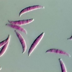
[ Morphology and physiology of fusarium
Fungi of the genus Fusarium form a well-developed mycelium of white, pink or red color. There are microconidia, macroconidia, rarely chlamydospores. Macroconidia are multicellular, spindle-sickle-shaped. Microconidia are oval, pear-shaped. They grow on Czapek medium in the form of fluffy colonies.
Pathogenesis and symptoms of fusarium
Fungi are widespread, especially on plants. In immunocompromised individuals, fungi can affect the skin, nails, cornea, and other tissues (F. moniliforme, F. sporotrichiella, F. anthapitum, F. chlantydosporum). Fever develops, rashes appear. The lesions are localized mainly on the extremities.
At low temperatures, the fungus F. sporotrichiella develops on cereals, producing mycotoxins. Eating such cereals that have overwintered under snow causes mycotoxicosis. Mycotoxicoses are also caused by eating grain products. The central nervous system is affected, with impaired coordination of movements.
Microbiological diagnostics of fusarium
Nails, skin, subcutaneous tissue, cornea, blood, the tip of a permanent catheter, vomit, feces, and tissue biopsies are examined. Fungi are isolated and their toxins are determined. RIF is used. Fluffy or cotton-like white colonies grow on nutrient media, which acquire a lilac-blue, pink-red, yellow, or green color as they age. Fungi form mycelium, micro- and macroconidia. Old cultures can form chlamydospores. PCR is sometimes used.