Medical expert of the article

New publications
Fasciola
Last reviewed: 04.07.2025

All iLive content is medically reviewed or fact checked to ensure as much factual accuracy as possible.
We have strict sourcing guidelines and only link to reputable media sites, academic research institutions and, whenever possible, medically peer reviewed studies. Note that the numbers in parentheses ([1], [2], etc.) are clickable links to these studies.
If you feel that any of our content is inaccurate, out-of-date, or otherwise questionable, please select it and press Ctrl + Enter.
Fasciola (common fasciola) is a flatworm from the trematode class. It affects livestock and causes weight loss, decreased milk yield and death of animals. Fascioliasis (a disease caused by liver fluke) occurs quite rarely in humans. The source of helminthiasis are animals infected with fasciola. The geography of the disease is quite extensive, from countries with a warm, humid climate (Peru, Chile, etc.) to territories with moderate climatic conditions (Belarus, France, etc.).
Structure fasciolas
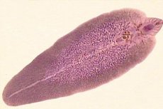
Fasciola has a flattened leaf-shaped body. The length fluctuates around 2-3 cm and the width up to 1 cm. It consists of two parts - a beak-shaped front part and a wide back part. The helminth has a pair of suckers of different sizes: the smaller one is the oral part, the larger one is the abdominal part.
The digestive tract of the common fasciola has a branched anatomical structure. At the beginning of the fasciola tract there is an oral sucker, which passes into the prepharyngeal cavity, then comes the pharynx and esophagus, which divides into branched intestinal loops. The fasciola is a hermaphrodite. In the middle part of its body are the testicles, here are also the ovaries, yolk glands and uterus. The shape of the fasciola eggs is oval with a two-layer yellowish membrane. The size of the eggs varies, starting from 120 microns in length.
Life cycle fasciolas
In its development, fasciola goes through several stages, accompanied by a change of hosts. Helminth eggs, released by the carrier into the external environment, enter the body of the intermediate host, where the formation and evolution of fasciola occurs. Developing, the larva reaches the body of the main host in different ways, where its final sexual maturation occurs. The intermediate hosts of fasciola are mollusks, snails, living in fresh water bodies.
The final host is a mammal (cattle and small cattle) or a human. Fasciola can live for several years in its bile ducts.
The parasite's host excretes helminth eggs into the environment with feces. When fertilized and viable fasciola eggs fall into water, they continue to develop. An aquatic environment with a temperature of 22 to 29 ° C is optimal for egg maturation and the emergence of larvae. Low (below +10 °C) and high (above +30° C) temperatures have a detrimental effect on the initial stage of fasciola development.
After 18 days, miracidia, adapted to life in water, emerge from the eggs. They penetrate the body of the intermediate host - a small freshwater snail. After 1-2.5 months, having passed the necessary stages of evolution, cercariae (tailed worms) appear. They leave the temporary host and again enter the water.
Using suckers, cercariae attach to the leaves of aquatic plants and emerge from the cyst. For greater survival, the larvae are covered with a dense shell. This period is called adolescaria - the appearance of larvae capable of invading the body of the main host. Adolescaria, in the presence of moisture, are well preserved (up to 1 year), but quickly die in a dry external environment (after 3 months). The body of the final host receives the larva together with contaminated water, fresh grass, poorly dried hay.
Adolescaria fasciola, having entered the intestinal lumen, penetrates the intestinal mucosa. From there, it penetrates the liver through the bloodstream and attaches to the bile ducts, where it begins to parasitize. With the bloodstream through the vessels, the larvae can reach atypical places of parasitism in the body - the lungs, mammary glands, skin. After 1.5-2 months from penetrating the body of the final host, fasciola turn into a sexually mature individual with a hermaphroditic reproductive system. Having achieved the formation and maturation of the reproductive system, fasciola is able to lay eggs. During the parasitization, fasciola lays up to 2 million eggs.
Pathogenesis
The method of introduction of fasciola is oral. It occurs exclusively with the use of raw untreated fresh water, unwashed greens, watered with water infected with cysts of the fluke. Eating insufficiently heat-treated goat or lamb liver infected with larvae of the fluke can lead to helminthic invasion.
Symptoms
The incubation period lasts from 1 week to 2 months. Infection can occur in two forms - acute and asymptomatic.
For the acute variant of the disease, the characteristic symptoms are allergic rashes (urticaria), weakness, a rise in body temperature to 39-40º C, cephalgia, pain in the epigastric region, in the right hypochondrium, nausea, vomiting, the appearance of yellowness of the skin, hepatomegaly, pain and compaction of the liver during palpation. From the cardiovascular system - the heart rate increases, the heart sounds are muffled, without rhythm disturbances, complaints of chest pain may be present. In the general blood test - a significant increase in the number of eosinophils, leukocytes, an increase in ESR of more than 20 mm / hour.
Asymptomatic stage. Begins 1.5-2 months after invasion. At this stage of the disease, symptoms of gastroduodenitis appear (decreased appetite, nausea periodically appears, abdominal pain of unclear localization, instability of stool - from diarrhea to constipation), attacks of spastic pain in the right hypochondrium, liver dysfunction are possible. In the biochemical parameters of the blood, the following are noted: increased values of ALT, AST, alkaline phosphatase, GGT, total bilirubin, abnormalities in the protein composition of the blood, decreased albumin values, an increase in gamma globulin levels. In the picture of the general detailed analysis of peripheral blood, an increase in eosinophils (up to 10%), slight anemia is recorded.
Diagnostics
The diagnosis is made based on clinical manifestations (a symptom complex of acute or asymptomatic manifestations of helminthic invasion), epidemiological history (bathing or drinking water from stagnant bodies of water, eating unwashed greens) and the results of diagnostic laboratory tests.
At an early stage of helminth infection, a stool smear test using the Kato method will not give an informative result, because the release of eggs by a mature helminth occurs 3-3.5 months after its entry and fixation in the liver ducts. At this stage, blood serum tests (RNGA, ELISA reactions) are of primary importance. In the case of an asymptomatic variant of helminthic invasion or suspected fascioliasis, a smear and stool test using the Kato method or analysis of the contents of the duodenum can be effective. It is possible to detect the presence of fasciola eggs in feces and the contents of the upper intestine. In the case of asymptomatic fascioliasis, it is impossible to determine exactly when the helminth entered the body and at what stage of sexual maturation it is. Feces analysis is carried out twice with an interval of 7-10 days.
Differential diagnosis
Fasciola invasion is differentiated from allergic conditions, gastroduodenitis, hepatitis, cholecystitis, cholangitis, helminthiasis caused by other representatives (opisthorchiasis, enterobiasis, taeniasis, clonorchiasis, trichinosis), etc.
The difference between pinworm and fasciola
Externally, the helminth fasciola is very different from pinworm. The symptoms of invasion can be similar. When pinworms enter the human body, they cause a disease called enterobiasis. Children often suffer from it. When the clinical picture of helminthic intoxication is not clearly expressed, intestinal symptoms are not significant, skin allergic reactions such as urticaria may appear. As with infection with the liver fluke, the introduction of pinworms provokes a state of sensitization of the body and manifestations of skin reactions. You have to seek help from a medical institution to immunologists-allergists. It is difficult to independently identify an allergen that provokes acute reactions of the immune system. When conducting laboratory tests aimed at determining the allergen, it is possible to identify helminthic invasion. In such cases, it is necessary to differentiate enterobiasis from invasion by the liver fluke.
The main differences are:
- Pinworms are a different type of helminth, they are of different sexes, only the female lays eggs;
- Invasion occurs when helminth eggs enter the digestive tract from dirty hands, unwashed vegetables and fruits;
- The localization site of pinworms is the large intestine. Here, individuals emerge from the cysts. After fertilization is complete, the female crawls to the anus and lays eggs, which causes itching and irritation in the anal area. This is the main distinguishing feature of pinworm infestation. To confirm or refute the diagnosis, an anal scraping is prescribed to determine the presence of eggs.
 [ 27 ], [ 28 ], [ 29 ], [ 30 ], [ 31 ], [ 32 ]
The difference between fasciola and bovine tapeworm
Beef tapeworm and fasciola have similar and distinctive features and belong to different types of helminths. Invasion by the tapeworm bovine tapeworm is possible when contaminated meat that has not been sufficiently cooked is ingested. Similarities are manifested in the structure of the worms and the way they attach to the body of the main host. The bovine tapeworm attaches to the intestines with the help of suckers and is a hermaphroditic representative of the species. The clinical picture at the onset of the disease is also similar - the presence of anemia, eosinophilia, leukocytosis, skin allergic reactions, weakness, nausea and vomiting. A feature of the bovine tapeworm is its enormous size (up to 5 meters) and the full cycle of sexual maturation and development occurs in the intestine. Its long-term presence in the human body leads to pathological loss of muscle and fat mass and severe intoxication. Throughout life, after sexual maturation of the individual, segments (segments) are separated from the helminth for the purpose of reproduction. They contain invasive larvae. The segments come out into the external environment through the anus, without causing itching.
The parasitic helminth is difficult to detect, diagnosis of the disease is difficult. In the absence of therapy, a person experiences pathological weight loss and suppression of immunity.
The main sign of the presence of bovine tapeworm in the body is the presence of segments in the feces.
Treatment
Hospitalization is advisable if there is a suspicion of liver fluke infestation and at the stage of early manifestations of the acute form of the disease. Outpatient treatment is possible.
At the early stages of the diagnosed disease, antiparasitic therapy is not prescribed, in order to avoid deterioration of the patient's health due to intoxication of the body with the decay products of the fasciola when it dies. At this stage of the disease, symptomatic and palliative treatment is prescribed. The choice of tactics and methods of treatment is decided by a helminthologist. The following drugs can be prescribed:
Enzyme-containing - mezym; kreazim; panzinorm; kreon; enzystal, etc.
Hepatoprotective and choleretic action - legalon; carsil; heptral; silegon; chophytol, etc.
Antihistamines - zodac; claritin; cetrine; diazolin; aerius, etc.
Affecting intestinal motility - duspatalin; sparex; niaspam; No-Spa, etc.
Probiotics - bifidum; florin forte; linex; bifiform etc.
Infusion therapy for the purpose of detoxifying the patient's body.
If indicated, broad-spectrum antibiotics and medications from other nosological groups are prescribed.
Antiparasitic therapy is indicated in the absence of symptoms characteristic of the acute form of the disease. Hexachlor-para-xylene (Chloxyl) is used - from 0.1 to 0.15 g / kg / day, biltricide (praziquantel) - 75 mg / kg. Therapy is carried out under careful medical supervision.
Follow-up tests should be performed regularly after 3 or 6 months of treatment.
Prevention fasciolas
To avoid infection with liver fluke, it is necessary to follow the rules:
- Avoid using unboiled standing water from ponds. If there is no alternative source of water and no possibility of boiling, it is necessary to filter it through a cloth.
- Be sure to wash greens (parsley, dill, cilantro, etc.) with water, then scald with boiling water or blanch in boiling water for several minutes.
- Conduct preventive deworming measures for cattle: feeding dry hay, prepared and aged for 6 months in storage, changing pastures, fighting snails in water bodies.
- Timely identification and deworming of patients with fascioliasis.