
Medical expert of the article

New publications
Computed tomography of the lungs: what the test shows, when it is prescribed, and how safe is it?
Last updated: 20.03.2026

All iLive content is medically reviewed or fact checked to ensure as much factual accuracy as possible.
We have strict sourcing guidelines and only link to reputable media sites, academic research institutions and, whenever possible, medically peer reviewed studies. Note that the numbers in parentheses ([1], [2], etc.) are clickable links to these studies.
If you feel that any of our content is inaccurate, out-of-date, or otherwise questionable, please select it and press Ctrl + Enter.
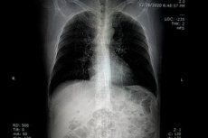
Computed tomography (CT) of the lungs is a method of layered imaging of the chest. An X-ray tube and detectors rotate around the body, and a computer assembles multiple thin slices into a detailed image of the lungs, pleura, mediastinum, major vessels, thoracic bones, and parts of the upper abdomen. Unlike conventional radiography, the doctor sees not a summary shadow of all structures at once, but rather thin anatomical layers that can be studied in different planes and even in 3D reconstruction. This is why CT scanning often helps clarify what appears unclear or suspicious on a conventional image. [1]
Modern CT scans of the lungs exist in more than one form, but several clinically distinct formats. There's standard chest imaging, low-dose CT for lung cancer screening, high-resolution CT for assessing interstitial and fibrosing lung diseases, and CT angiography for vascular imaging, such as pulmonary embolism. Not only the technical settings but also the diagnostic purpose vary. [2]
The main advantage of this method is that it simultaneously accurately depicts lung tissue, pleura, bone, soft tissue, and vascular structures. Radiological societies emphasize that CT produces much more detailed images than traditional radiography, and its high scanning speed makes it particularly useful in emergency medicine, cases of severe shortness of breath, trauma, and situations where it is necessary to quickly understand what is happening inside the chest. [3]
In everyday practice, CT scanning of the lungs is ordered not because it is "the best overall scan," but because it answers a specific clinical question. It is used to clarify changes detected on radiography, to find the cause of cough, shortness of breath, chest pain, or fever, to evaluate tumors and their extent, to monitor the response to treatment, to evaluate trauma, and to diagnose a wide range of lung diseases—from pneumonia and bronchiectasis to interstitial lesions and congenital anomalies. [4]
However, there is no universal template for the examination. If an answer is needed about minor changes in the lung parenchyma, a non-contrast option may be optimal. If the goal is to evaluate vessels, hemoptysis, the pleura, or tumor spread to adjacent structures, contrast enhancement or computed tomography angiography is often required. Therefore, the correct examination is not simply a matter of "taking a CT scan," but rather selecting a protocol tailored to the task. [5]
Source of criteria for section and table: [6]
| Research option | The main goal | Contrast |
|---|---|---|
| Standard chest computed tomography | Clarification of foci, inflammation, tumors, trauma, pleural changes | According to the readings |
| Low-dose computed tomography | Lung cancer screening in high-risk groups | Usually without intravenous contrast |
| High-resolution computed tomography | Interstitial diseases, fibrosis, small parenchymal changes | Usually without intravenous contrast |
| Computed tomography angiography | Evaluation of the thoracic vessels, especially if a pulmonary artery thrombus is suspected | An iodinated contrast agent is required. |
| Contrast-enhanced computed tomography of the chest | Tumors, mediastinum, hemoptysis, part of pleural and vascular problems | Required for a clinical question |
When is a CT scan of the lungs really necessary?
Computed tomography (CT) of the lungs is especially useful when conventional imaging is insufficient. If an X-ray reveals a densification, nodule, infiltrate, suspected tumor, or unexplained lesion, CT is often the next logical step. The same applies to situations where a patient has chest symptoms, but a conventional X-ray cannot reliably determine the cause of cough, shortness of breath, chest pain, or fever. [7]
For chronic shortness of breath, CT scanning is not prescribed equally for everyone. The American College of Radiology recommends that if chronic obstructive pulmonary disease, small airway disease, or complications from COVID-19 are suspected, a chest CT scan without contrast is often appropriate. This is important because many patients mistakenly believe that intravenous contrast is essential for any lung examination, although in some clinical scenarios it provides no additional benefit. [8]
With hemoptysis, the situation changes. The American College of Radiology criteria for non-massive and recurrent hemoptysis list contrast-enhanced computed tomography and chest computed tomography angiography as appropriate initial tests, as the physician must evaluate not only the lung tissue but also the vascular structures, the source of bleeding, and possible tumor or inflammatory causes. Therefore, the choice of protocol depends directly on the symptom. [9]
In interstitial lung diseases and early pulmonary fibrosis, the value of computed tomography is particularly significant. The 2025 American Thoracic Society Clinical Position Statement emphasizes that computed tomography is used to detect interstitial abnormalities and helps differentiate early changes from established interstitial lung disease. An editorial in the same publication specifically notes that high-resolution computed tomography remains the standard imaging method for assessing interstitial lung disease. [10]
When pulmonary embolism is suspected, computed tomography angiography plays a key role. European guidelines on pulmonary embolism indicate that multislice computed tomography angiography has become the method of choice for visualizing the pulmonary vasculature in patients with suspected pulmonary embolism. This is one of the best examples of how "conventional lung tomography" and a vascular protocol are essentially different studies with different objectives. [11]
Source of criteria for section and table: [12]
| Clinical situation | What does a doctor most often need? | Which research option is more appropriate? |
|---|---|---|
| Unclear change on radiography | To clarify the nature and distribution of the find | Standard computed tomography |
| Chronic shortness of breath, suspected chronic obstructive pulmonary disease | Assess the parenchyma and airway | More often without contrast |
| Suspected small focal interstitial changes | Assess lung tissue in the most detailed possible way | High-resolution computed tomography |
| Hemoptysis | Find the source of bleeding and evaluate the vessels | Contrast tomography or computed tomography angiography |
| Suspected thrombus in the pulmonary artery | Assess the pulmonary vascular bed | Computed tomography angiography |
| Lung cancer screening in high-risk groups | Find asymptomatic early lesions | Low-dose computed tomography |
How to prepare and how the study is conducted
Preparation depends primarily on whether intravenous contrast will be used. For most examinations, patients are advised to wear comfortable clothing and remove metal objects, jewelry, eyeglasses, removable dental appliances, and anything else that could impair image quality. This may seem like a small detail, but it is precisely these details that sometimes create artifacts and interfere with accurate image interpretation. [13]
If a contrast injection is planned, the patient may be asked to fast for several hours before the examination. Before the procedure, it is important to inform the doctor about all medications, allergies, recent illnesses, as well as any history of asthma, heart disease, diabetes, kidney disease, or thyroid disease. This information is not just a formality, but rather a genuine risk assessment and the selection of a safe protocol. [14]
The machine itself typically consists of a short, wide, ring-shaped tunnel with a narrow table running through it. The X-ray tube and detectors rotate inside, and the operator is located in an adjacent room, but constantly hears and sees the patient through an intercom. For those who fear the long, closed tunnels of magnetic resonance imaging, this is an important detail: CT scans are usually easier to tolerate precisely because of the machine's short design and very short scanning time. [15]
During the examination, the patient is positioned on a table, sometimes secured with pillows or belts to prevent unnecessary movement. If contrast is needed, it is injected into a vein immediately before the scan begins. The table then passes through the machine's ring several times, and the patient may be asked to briefly hold their breath. The scan itself typically takes less than 30 seconds, while the entire visit, including preparation, typically takes approximately 30 minutes. [16]
After the examination is completed, the intravenous catheter is removed, and in most cases, normal activities can be resumed immediately. The images are analyzed by a radiologist, who provides an official report for the attending physician. A follow-up examination is sometimes required after a period of time to assess the progress of the lesion, check its response to treatment, or determine whether the suspicious lesion has changed compared to previous images. [17]
Source of criteria for section and table: [18]
| Stage | What's happening | What is important for the patient |
|---|---|---|
| Before the study | Complaints, medications, allergies, kidney diseases, and the likelihood of pregnancy are clarified. | Tell your doctor all the important information |
| Preparation | Metal objects are removed, sometimes a fasting break is required | Do not hide implants, allergies, or medications. |
| Laying on the table | The patient is positioned correctly | Lie still |
| Scanning | The machine makes a series of thin slices, sometimes with a breath hold | Do not move, listen to staff commands |
| After research | The catheter is removed, the images are used for description. | You can usually return to your normal activities immediately. |
What does a CT scan of the lungs show and how are the results interpreted?
Computed tomography (CT) is effective in detecting focal and diffuse changes in lung tissue. It can reveal nodules and tumors, inflammatory infiltrates, areas of tuberculous lesions, bronchiectasis, pleural changes, congenital anomalies, and many other conditions. Radiological societies explicitly state that this method is routinely used to evaluate pneumonia, interstitial lung diseases, tumors, and other acquired or congenital disorders. [19]
In chronic and fibrosing lung diseases, CT scanning is particularly valuable because it allows for the visualization of reticular changes, areas of ground-glass consolidation, traction bronchiectasis, disruption of lung tissue architecture, and other features that are difficult or impossible to assess with conventional imaging. In the 2025 American Thoracic Society clinical statement, CT scanning is the basis for identifying interstitial abnormalities, non-fibrotic and fibrotic variants, and for monitoring their progression. [20]
In addition to lung tissue, the physician evaluates blood vessels, lymph nodes, the heart, large vessels, the pleural cavity, bones, and even part of the upper abdomen. A review of the screening report structure by RadiologyInfo emphasizes that the radiologist pays attention not only to pulmonary nodules but also to emphysema, coronary artery calcification, fluid around the lungs, the airway lumen, lymph nodes, and incidental findings in the bones and upper abdomen. This is why a lung CT scan often provides the physician with more information than the patient expected. [21]
Comparison with previous studies is extremely important. The same source explains that the comparison section in the report allows one to determine whether the lesion is new, growing, stable, or long-standing without dynamics. This is especially important for pulmonary nodules, because the same lesion, when it shows no growth during observation, and when it rapidly enlarges, are two completely different clinical situations. [22]
A typical report contains several key sections: the clinical history, the examination technique, a comparison with previous images, a description of the observed changes, and a summary in which the radiologist formulates the most important findings and recommendations. For screening low-dose imaging, a standardized outcome category system is additionally used to determine whether routine annual follow-up, an earlier repeat examination, a consultation with a pulmonologist, or a biopsy is needed. The American College of Radiology maintains a current standardized reporting system for screening, and RadiologyInfo explains to patients how these categories influence subsequent management. [23]
Source of criteria for section and table: [24]
| A tomography finding | What could this mean? |
|---|---|
| Pulmonary node | Benign nodule, scar, inflammation, early lung cancer |
| Frosted glass type area | Inflammation, interstitial lesion, part of the adenocarcinoma spectrum |
| Emphysema | Destruction of the alveoli, often associated with smoking |
| Traction bronchiectasis and subpleural reticular changes | Possible pulmonary fibrosis or interstitial disease |
| Pleural effusion | Fluid around the lung due to infection, heart failure, tumor, and other causes |
| Enlarged lymph nodes | Inflammation, infection, tumor process |
| Coronary artery calcification | Atherosclerotic lesions of the heart vessels |
| Thrombus in the branches of the pulmonary artery | Pulmonary embolism |
Benefits, limitations and risks of the method
The advantages of computed tomography of the lungs are well-established. The method is fast, widely available, painless, non-invasive, and accurate. It simultaneously displays bone, soft tissue, blood vessels, and lung tissue, aids in rapid decision-making in emergency situations, and, if necessary, is used to guide a biopsy needle. According to RadiologyInfo, it is this combination of speed, detail, and practical accessibility that makes it one of the most valuable chest imaging techniques. [25]
The primary risk that patients most often worry about is radiation exposure. According to RadiologyInfo, the estimated effective dose for a standard chest CT scan is approximately 6.1 millisieverts, and for low-dose CT used in lung cancer screening, it is approximately 1.5 millisieverts. For comparison, a conventional chest X-ray delivers approximately 0.1 millisieverts. This does not mean that the scan itself is dangerous, but it explains why a physician should order it based on indications, not just "just in case." [26]
Modern protocols actively reduce radiation dose. RadiologyInfo notes that low-dose CT can provide diagnostically adequate image quality with significantly lower radiation exposure, and in some situations, the dose reduction compared to conventional CT can be 65% or more. A separate patient resource on lung cancer screening states that low-dose CT can use up to 90% less radiation than conventional chest CT, if the examination objective allows for this mode. [27]
Contrast risks are real, but generally low and well-managed. The American College of Radiology's 2025 Guidelines for Contrast Agents state that allergic-like reactions to modern iodinated contrast agents are uncommon, with a combined incidence of approximately 0.6% and severe reactions occurring at approximately 0.04%. This means that the risk exists, but it is small, and the entire examination preparation system is designed to identify risk factors in advance, weigh the benefits and harms, and be prepared to promptly treat any potential reaction. [28]
Kidney function and pregnancy are a separate issue. RadiologyInfo points out that when using iodinated contrast, the physician considers the presence of chronic or acute kidney disease, and in cases of reduced kidney function, the contrast may further worsen it. It also emphasizes that CT scans are generally avoided for pregnant women unless absolutely necessary due to the potential risk to the fetus. This is not an absolute prohibition, but rather a reason to carefully justify the examination and choose the safest option. [29]
There are also purely technical limitations. A very large patient may not fit within the standard machine's aperture or exceed the table's load capacity. Body movement and breathing can blur the image. Furthermore, RadiologyInfo notes that some types of soft tissue abnormalities are better visible on magnetic resonance imaging. Therefore, CT is not the answer to every question, although it remains one of the most important techniques for the lungs and chest organs. [30]
Source of criteria for section and table: [31]
| Parameter | What is important to know |
|---|---|
| Speed | The shooting itself usually lasts less than 30 seconds. |
| Informativeness | The lungs, vessels, pleura, bones, and mediastinum are clearly visible. |
| Standard radiation exposure | About 6.1 millisieverts for a routine chest CT scan |
| Screening radiation exposure | About 1.5 millisieverts for low-dose screening |
| Contrast reactions | Rare, but require risk assessment in advance |
| Pregnancy | They try not to prescribe the study without a serious need. |
| Restrictions | Movements, large body mass, and some soft tissue tasks are better seen on magnetic resonance imaging |
Low-dose computed tomography in lung cancer screening
Low-dose computed tomography (LDCT) occupies a special place as a lung cancer screening tool. Here, the test is performed not because there are already complaints, but to detect early, asymptomatic cancer in people at high risk. This is the fundamental difference between screening and diagnostic CT. Symptomatic individuals require individualized diagnostic testing, while asymptomatic individuals from a high-risk group require a regular early detection program. [32]
Screening eligibility criteria vary among professional organizations. The U.S. Preventive Services Task Force recommends annual low-dose CT scanning for people aged 50–80 years with at least 20 pack-years of smoking history if they continue to smoke or have quit for less than 15 years. The American Cancer Society, in updated guidelines, also recommends annual screening for people aged 50–80 years with at least 20 pack-years of smoking history who are current or former smokers and emphasizes the need to discuss the benefits, limitations, and potential harms before initiating a screening program. [33]
This strategy works not only in theory. The National Lung Cancer Screening Study showed a 20% reduction in lung cancer mortality with low-dose CT scanning compared to chest X-ray, and the European NELSON study also showed a statistically significant reduction in mortality in high-risk individuals. It is these data that underpin current screening recommendations. [34]
After a screening CT scan, not only the finding itself is important, but also its standardized interpretation. RadiologyInfo explains to patients that a typical screening report describes the clinical history, imaging technique, comparison with previous images, findings, and a final conclusion. Suspicious nodules are categorized into outcome categories: low categories typically indicate a return for annual follow-up, intermediate categories indicate an earlier repeat scan, and high categories indicate the need for consultation and, sometimes, biopsy. The American College of Radiology maintains a current standardized reporting system that helps make decisions more consistent and safe. [35]
A new and important layer of evidence is that screening CT scans can detect more than just lung cancer. The American Thoracic Society's 2025 clinical statement recommends systematically recording the presence or absence of interstitial anomalies and interstitial lung disease in smokers undergoing low-dose screening. In other words, properly performed and accurately reported CT scans can become a window into the early detection of several serious chest diseases. [36]
Source of criteria for section and table: [37]
| Screening parameter | What is important to know |
|---|---|
| Target | Find early lung cancer before symptoms appear |
| Method | Annual low-dose computed tomography |
| The core high-risk group according to US recommendations | Age 50-80 years and smoking history of at least 20 pack-years |
| Additional condition in the U.S. Preventive Services Task Force recommendations | Continues to smoke or quit less than 15 years ago |
| Practical logic | Screening is not necessary for everyone, but only for high-risk groups |
| The strength of the method | Reducing mortality through earlier detection |
| What else can you notice? | Emphysema, coronary calcification, interstitial anomalies, other incidental findings |
FAQ
How does a CT scan of the lungs differ from a conventional chest X-ray?
A chest X-ray provides a comprehensive image of all chest structures in one or more projections, while a CT scan creates thin, layered sections and allows for a much more detailed view of lung tissue, the pleura, blood vessels, and the mediastinum. This is why a CT scan is often prescribed to clarify findings on a conventional X-ray, rather than as a literal replacement. [38]
Is contrast always necessary for lung tomography?
No. For many cases, especially when assessing chronic dyspnea, emphysema, small airways, interstitial changes, and some post-viral conditions, a non-contrast examination is sufficient. Contrast is especially necessary for vascular issues, hemoptysis, and some tumor and pleural issues. [39]
Can CT scans differentiate inflammation from cancer?
Sometimes yes, but not always. CT scans are very good at showing the shape, density, and edges of a lesion, as well as its connection to the bronchi, pleura, and blood vessels. However, some findings remain unclear and require comparison with previous images, a repeat examination over time, additional methods, or a biopsy. [40]
How long does the examination take?
The scan itself typically takes less than 30 seconds, and the entire process, including preparation, typically lasts about 30 minutes. This time may be extended if intravenous contrast, an additional series of images, or post-contrast monitoring is required. [41]
How much radiation exposure is involved?
A standard chest CT scan typically delivers about 6.1 millisieverts, while a low-dose screening CT scan delivers about 1.5 millisieverts. This is higher than that of a conventional chest X-ray, so the examination must be justified and the protocol optimized for the clinical need. [42]
Is iodinated contrast material dangerous?
In most cases, it is well tolerated. According to the American College of Radiology guidelines, severe allergic-like reactions are rare, but the risk cannot be completely ruled out. Therefore, a medical history is collected before the examination, kidney disease is assessed, and the department's preparedness to treat a possible reaction is assessed. [43]
Can a CT scan be performed during pregnancy?
This test is generally avoided unless absolutely necessary. If the issue is clinically important and the mother's safety depends on it, the scan may be performed, but only after an individual assessment of the benefits and risks. [44]
What should you do if the report mentions a "nodule," "ground glass opacity," or an "incidental finding"?
First of all, don't interpret this as a diagnosis on your own. RadiologyInfo emphasizes that it's the report section that determines what the treating physician should focus on and whether observation, earlier follow-up, specialist consultation, or biopsy is required. For some nodules, dynamic observation is sufficient, especially if old images are available for comparison. [45]
Key points from experts
Ella Kazerooni, MD, MS, professor of radiology and internal medicine at the University of Michigan, is chair of the American College of Radiology's first committee on a standardized lung cancer screening system. A key expert thesis from her line of work is that screening low-dose CT should not just be an image, but part of a standardized quality system where outcome categories are directly linked to a patient's safe path forward. [46]
Anna Podolanchuk, MD, MS, a pulmonologist and critical care physician, is a recognized expert in interstitial lung diseases. The key message of the 2025 American Thoracic Society position statement, of which she is the co-lead author, is that chest imaging today serves not only to confirm advanced fibrosis but also to facilitate early recognition of interstitial abnormalities, their risk stratification, and surveillance planning. [47]
Matthew Davenport, MD, MBA, professor of radiology and a recognized expert on contrast agent safety, explains: "The rationale behind the American College of Radiology's contrast agent guidelines, developed by a committee, is simple and very practical: before administering a contrast agent, one should not automatically fear it or ignore the risks, but rather weigh the diagnostic benefits, alternatives, patient history, and the willingness to quickly recognize and treat rare adverse reactions. [48]
Stavros Konstantinidis, MD, Professor of Clinical Research and Medical Director of the Center for Thrombosis and Hemostasis at the University of Mainz, is one of Europe's leading experts on pulmonary embolism. His research suggests that computed tomography angiography plays a central role in suspected pulmonary embolism because the vascular protocol allows for rapid and reliable assessment of the pulmonary arterial bed. This is especially important for patients who believe that all lung CT scans are the same. [49]