Medical expert of the article

New publications
Angiography of the maxillofacial region
Last reviewed: 04.07.2025

All iLive content is medically reviewed or fact checked to ensure as much factual accuracy as possible.
We have strict sourcing guidelines and only link to reputable media sites, academic research institutions and, whenever possible, medically peer reviewed studies. Note that the numbers in parentheses ([1], [2], etc.) are clickable links to these studies.
If you feel that any of our content is inaccurate, out-of-date, or otherwise questionable, please select it and press Ctrl + Enter.
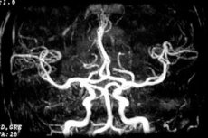
Angiography is a contrast X-ray examination of the arterial system (arteriography) and veins (venography) of the maxillofacial region - it is carried out by:
- percutaneous puncture of the external carotid artery;
- retrograde catheterization of the external carotid artery through the superficial temporal or facial artery;
- percutaneous catheterization using the Seldinger technique through the femoral or common carotid artery with the introduction of a catheter into the external carotid artery.
After catheterization of the external carotid artery or one of its branches (selective angiography), 5 to 15 ml of contrast agent (verografin, urografin, omnipaque, etc.) is injected into the vessel and a series of images are taken.
Angiography is used to diagnose anomalies and diseases of the vascular system (hemangiomas, juvenile angiofibromas of the skull base); X-ray vascular embolization is also performed in the treatment of hemangiomas of the maxillofacial region.
Contraindications to the study include hypersensitivity to iodine, active pulmonary tuberculosis, liver and kidney damage, recent myocardial infarction and stroke, and the general severe condition of the patient.
Direct lymphography is performed after the introduction of a dye into the thickness of the skin to visualize the lymphatic vessels. An incision to locate the vessel for cosmetic reasons is made in the area behind the ear and ultra-liquid lipoiodol is introduced into the lymphatic vessel taken on the ligature. The radiographs show the lymphatic vessels and contrasted regional lymph nodes.
 [
[