Medical expert of the article

New publications
Test for TTH in pregnancy in the 1st, 2nd and 3rd trimester: deciphering the indicators
Last reviewed: 05.07.2025

All iLive content is medically reviewed or fact checked to ensure as much factual accuracy as possible.
We have strict sourcing guidelines and only link to reputable media sites, academic research institutions and, whenever possible, medically peer reviewed studies. Note that the numbers in parentheses ([1], [2], etc.) are clickable links to these studies.
If you feel that any of our content is inaccurate, out-of-date, or otherwise questionable, please select it and press Ctrl + Enter.
TSH during pregnancy may have different values than under normal conditions. Monitoring thyroid function is very important for both healthy women and women with existing thyroid dysfunction. After all, the child's development depends on the function of many organs of the woman, including the thyroid gland.
Indications for the procedure T.T.H. tests in pregnancy
Indications for screening the TSH level are the appearance of symptoms that are characteristic of hypothyroidism - drowsiness, inadequate weight gain, the appearance of dense edema, and skin trophic disorders. If there are such symptoms, then we are talking about the clinical form of hypothyroidism in pregnant women, which means that examination in such cases is mandatory. But what to do if the course of hypothyroidism is subclinical. In such cases, if a woman wants to give birth to a healthy child, methods of planning a healthy pregnancy should come to the fore. Pre-pregnancy examinations of the mother in such cases should also include screening of the thyroid gland function.
TSH when planning a pregnancy can become a screening test that will help determine whether a woman has any disorders. The TSH norm when planning a pregnancy should be within 0.4–4.0 mIU/L. If a woman has problems with the thyroid gland or is undergoing treatment for thyroid pathology, the TSH level when planning a pregnancy should not exceed 2.5 mIU/L. This level will allow the embryo to implant normally and develop normally.
Preparation
There are no special instructions for preparing for this test. It is not recommended to consume alcohol, nicotine, or medications the day before the test. If a woman takes thyroxine or other medications to treat thyroid function, she should stop taking them the day before.
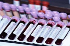
How to take TSH during pregnancy? This is done in the laboratory in the morning on an empty stomach. Venous blood is taken with subsequent testing over several days.
Who to contact?
Technique T.T.H. tests in pregnancy
Determination of serum or plasma levels of thyroid stimulating hormone (TSH) is recognized as a sensitive method in the diagnosis of primary and secondary hypothyroidism. TSH is secreted by the anterior pituitary gland and stimulates the production and release of thyroxine and triiodothyronine by the thyroid gland. Although the concentration of TSH in the blood is extremely low, it is sufficient to maintain normal thyroid function. The release of TSH is regulated by TSH-Releasing Hormone (TRH), produced by the hypothalamus. The levels of TSH and TRH are inversely related to the level of thyroid hormones. When there is a high level of thyroid hormones in the blood, less TRH is released by the hypothalamus, so that less TSH is released by the pituitary gland. The opposite effect will occur when there is a decrease in thyroid hormones in the blood. This process is known as the negative feedback mechanism and is responsible for maintaining proper levels of these hormones in the blood.
Normal performance
The norm of TSH during pregnancy varies by trimester, which is due to different levels of T3 and T4 synthesis throughout pregnancy. Different indicators may differ in different laboratories, but there are average recommended indicators of TSH levels at different stages:
- TSH during pregnancy in the first trimester should be within the range of 0.1 – 2.5 mIU/L;
- TSH during pregnancy in the 2nd trimester should be within the range of 0.2 – 3.0 mIU/L;
- TSH during pregnancy in the 3rd trimester should be within the range of 0.2 – 3.5 mIU/L.
If there are deviations of any values, then a comprehensive assessment of the thyroid gland function is carried out. For this, the levels of TSH, T3 and T4 during pregnancy are examined, which can indicate one or another function of the thyroid gland.
The device for analysis
The TSH assay uses a monoclonal antibody. The ELISA kit is used to quantify the concentration of thyroid stimulating hormone (TSH) in human serum. This TSH kit is based on the enzyme-linked immunosorbent assay principle. It uses a unique monoclonal antibody directed against a distinct antigenic determinant on the intact TSH molecule. A mouse monoclonal anti-TSH antibody is used to immobilize the solid phase (wells on a microtiter plate). A goat anti-TSH antibody is suspended in an enzyme conjugate solution. The test sample reacts simultaneously with these two antibodies, resulting in TSH molecules being sandwiched between the solid phase and the enzyme-linked antibodies. After 60 minutes of incubation at room temperature, the wells are washed with water to remove unbound labeled antibodies. TMB solution is added and incubated for 20 minutes, which results in the development of a blue color. The color development is stopped by adding stop solution, which results in the formation of a yellow color, and the measurement is carried out on a spectrophotometer at a wavelength of 450 nm. The concentration of TSH is directly proportional to the color intensity of the sample. The minimum detectable concentration of TSH with this kit is 0.2 μIU/ml.
Raising and lowering of values
Elevated TSH during pregnancy is one of the laboratory signs of hypothyroidism in the woman, and therefore of hormone deficiency in the child. Elevated TSH with normal T4 and T3 concentrations is defined as subclinical hypothyroidism. The prevalence of subclinical hypothyroidism during pregnancy is estimated at 2% to 5%. It is almost always asymptomatic. Women with subclinical hypothyroidism are more likely than euthyroid women to have positive TPO antibody activity. Subclinical hypothyroidism is associated with poor maternal and offspring outcomes, and most recommend thyroxine replacement in women with subclinical hypothyroidism. However, although thyroxine treatment improves obstetric outcome, it has not been shown to alter long-term neurodevelopmental outcomes in the offspring. The consequences of high TSH for the child are not limited to low birth weight. The child may be born with features of congenital hypothyroidism. This pathology is characterized by insufficient development of internal organs, and mainly connections in the brain. If congenital hypothyroidism is not diagnosed, the child develops a profound cognitive neurological deficit.
High TSH and a frozen pregnancy may have a direct connection. Since thyroid hormones support pregnancy by stimulating the function of the corpus luteum, their deficiency may cause a frozen pregnancy.
How to lower TSH during pregnancy if its increase is dangerous. First of all, it is important to understand that we cannot directly influence TSH synthesis through medications. If TSH is elevated in the body, this only indicates that the level of T3 and T4 is below normal. Therefore, it is necessary to increase the concentration of these hormones, and TSH will increase accordingly. If there is low T3 and T4 against the background of high TSH, then the hormone thyroxine is necessarily used in treatment. The introduction of levothyroxine is the treatment of choice for maternal hypothyroidism. Pregnant women need high doses due to the rapid increase in TSH levels as a result of the physiological increase in estrogen, increased placental transport and metabolism of maternal T4, and an increase in the volume of distribution of thyroid hormones. During pregnancy, the complete replacement dose of thyroxine is about 2-2.4 mcg / kg / day. In severe hypothyroidism, a thyroxine dose up to twice the expected final replacement daily dose may be given for the first few days to rapidly normalize the extrathyroidal thyroxine pool before the final replacement dose is reduced. Women already on thyroxine before pregnancy typically need to increase their daily dose by an average of 30% to 50% above the preconception dose. The thyroxine dose also depends on the etiology of the hypothyroidism. Women should have T4 and TSH values monitored every 4 to 6 weeks until delivery.
Maternal dietary iodine deficiency results in impaired thyroid hormone synthesis in the mother and fetus. Low thyroid hormone levels stimulate increased pituitary TSH production, and elevated TSH stimulates thyroid growth, leading to maternal and fetal goiter. Therefore, elevated TSH may not be due to low T3 and T4 levels but may be primarily due to iodine deficiency. In areas with severe iodine deficiency, thyroid nodules may be present in up to 30% of pregnant women. Severe iodine deficiency in pregnant women is associated with increased rates of pregnancy loss, stillbirth, and increased perinatal and infant mortality.
Normal levels of thyroid hormones are necessary for neuronal migration, myelination, and other structural changes in the fetal brain. Because thyroid hormones are needed throughout pregnancy, iodine deficiency affects both maternal and fetal thyroid hormone production, and inadequate iodine intake can have deleterious effects. In particular, maternal and fetal iodine deficiency during pregnancy negatively affects the cognitive function of the offspring. Children whose mothers were severely iodine deficient during pregnancy may exhibit cretinism, characterized by profound intellectual disabilities, deafness, and motor impairment. Iodine deficiency is the leading cause of preventable intellectual disabilities worldwide.
In such cases, the use of levothyroxine to increase the level of T3 and T4 and to decrease TSH is inappropriate; it is necessary to first correct the level of iodine deficiency. Iodomarin with increased TSH during pregnancy is in this case the drug of choice for the treatment of iodine deficiency. All pregnant and lactating women with this problem need to take Iodomarin, which contains 150-200 mcg of iodine per day.
Hyperthyroidism is less common than hypothyroidism, with an estimated incidence of 0.2% during pregnancy. Low TSH in pregnancy and elevated T4 are laboratory signs of hyperthyroidism in women. Sometimes there is a low TSH with normal T4 in pregnancy, which is characteristic of subclinical hyperthyroidism. Clinical symptoms of hyperthyroidism include tachycardia, nervousness, tremor, sweating, heat intolerance, proximal muscle weakness, frequent bowel movements, decreased exercise tolerance, and hypertension.
The reasons for such changes are the formation of an autoimmune process. In this pathology, antibodies (Ab) to TSH receptors are formed, which are elevated during pregnancy precisely in the case of hyperthyroidism. These antibodies stimulate the production of TSH in a false way, which in turn stimulates the production of thyroid hormones. These hormones increase in the blood and lead to the activation of all functions of the thyroid gland and other organs and systems of the pregnant woman.
The main concern in women with hyperthyroidism is the potential impact on the fetus. Thyroid receptor antibodies should be measured by the end of the second trimester in women with active disease.
Changes in thyroid function during pregnancy
Pregnancy is a period that at the best of times creates great physiological stress for both mother and fetus. However, when pregnancy is complicated by endocrine disorders such as hypothyroidism, the potential for adverse maternal and fetal outcomes can be enormous. Hypothyroidism is common among pregnant women, and the rate of detection, especially in developing countries, has kept pace with the magnitude of the problem. Because hypothyroidism is easily treated, early detection and treatment of the disorder can reduce the burden of adverse fetal and maternal outcomes that are very common.
Thyroid dysfunction during pregnancy is common, with an incidence of 2%-4%. Maternal thyroid dysfunction is associated with an increased risk of a variety of adverse maternal and child outcomes, including miscarriage, intrauterine growth restriction, hypertensive disorders, preterm birth, and decreased IQ in the child. During pregnancy, profound changes in thyroid physiology occur to ensure adequate thyroid hormone levels for both the mother and the fetus. This is especially important during early pregnancy because the fetal thyroid gland does not begin to produce significant amounts of TSH until approximately 20 weeks of gestation, until which time the fetus is highly dependent on maternal hormone levels. This suppression of fetal thyroid hormone synthesis, as well as increased concentrations of hormone binding proteins (thyroxine-binding globulin) and degradation of T4 by placental iodothyronine deiodase 3, necessitate increased maternal thyroid hormone production. This requires a healthy maternal thyroid gland and adequate availability of dietary iodine. As a consequence, serum free thyroxine (FT4) concentrations increase and TSH concentrations decrease from about the eighth week through the first half of pregnancy, resulting in different reference intervals for TSH and T4 compared with the non-pregnant state.
Given these pregnancy-related changes in thyroid physiology and complications associated with thyroid dysfunction, it is important to establish reference intervals for normal thyroid function during pregnancy. This is critical to identifying women who require treatment or correction of thyroid function.
Thyroid dysfunction that is not diagnosed in time can be a problem. While much attention has been focused on the adverse fetal outcomes associated with hypothyroidism, attention is also gradually being directed to the adverse maternal outcomes of this disorder. Prompt diagnosis and treatment of hypothyroidism during pregnancy are very important. Subclinical hypothyroidism also needs to be identified and treated to prevent adverse outcomes, especially maternal. Since women with hypothyroidism during pregnancy, especially the autoimmune variety, may have a relapse of the disorder after delivery or may continue to require thyroxine replacement after delivery, adequate follow-up is essential. And even if a woman was completely healthy before pregnancy and never had thyroid disorders, such problems may appear even in the context of a normal pregnancy.
Thyroid physiology undergoes marked changes during normal pregnancy. These changes occur throughout pregnancy, help prepare the maternal thyroid to cope with the metabolic demands of pregnancy, and are reversible after delivery.
The most notable change is an increase in thyroxine-binding globulin (TBG). This begins early in the first trimester, plateaus during midlife, and persists until delivery. This is due to stimulation of TBG synthesis by elevated maternal estrogen levels and, more importantly, to decreased hepatic clearance of TBG due to estrogen-induced sialylation. This increased TBG concentration results in pool expansion and leads to increased total T3 and T4 levels due to increased maternal thyroid hormone synthesis. Maternal thyroid hormone synthesis is also increased by accelerated renal clearance of iodide as a result of increased glomerular filtration rate.
Increased T4 metabolism in the second and third trimesters, due to increased placental type II and type III deiodinases, which convert T4 to T3 and T4 back to T3 and T2, respectively, act as an additional stimulus for T4 synthesis. Plasma iodide levels decrease due to increased thyroxine metabolism and increased renal iodide clearance. All these changes result in an increase in thyroid size in 15% of pregnant women, which returns to normal in the postpartum period.
Serum hCG has its own thyroid-stimulating activity, which increases after fertilization and peaks at 10-12 weeks. Consequently, free T3 and T4 levels increase slightly in the first trimester, and TSH levels decrease in the first trimester, with correction in the second and third trimesters when hCG levels decrease.
How does TSH affect pregnancy? Given that its level slightly decreases according to the feedback principle in the first trimester, its effect also decreases slightly. But the synthesis of this hormone is preserved, and it affects not only the woman's body itself, but also the thyroid gland in the child, which is actively developing.
The fetal thyroid gland develops until 7 weeks of gestation. The fetal gland is able to take up iodine by 12 weeks and can synthesize thyroxine by 14 weeks of gestation. However, significant hormone secretion does not occur until 18–20 weeks of gestation. Thereafter, fetal TSH, T4, and TSH gradually increase to adult levels by 36 weeks of gestation. Transplacental TSH transport is negligible, but T3 and T4 transport may be significant.
Thus, it can be concluded that the mother's thyroid gland performs functions for the fetus up to a certain period of pregnancy. Therefore, the mother herself may experience various thyroid deficiencies, especially if she previously had hypothyroidism or hyperthyroidism. Monitoring thyroid function during pregnancy is very important, because even clinically unnoticeable hypothyroidism in the mother can cause serious cognitive impairment and organ development disorders in the child.
 [ 23 ], [ 24 ], [ 25 ], [ 26 ]
Monitoring thyroid function during pregnancy
Undiagnosed maternal hypothyroidism may result in preterm birth, low birth weight, and respiratory distress in neonates. Over the years, ample evidence has accumulated regarding the role of thyroxine in normal fetal brain development. The presence of specific nuclear receptors and thyroid hormones detected in the fetal brain at 8 weeks of gestation, free T4 detected in coelomic and amniotic fluids, and the demonstration of placental transfer of maternal thyroid hormones highlight the role of thyroid hormones in fetal brain development. Complex interactions between iodothyronine deiodases D2 and D3 during pregnancy help fine-tune the amount of adequate T3 required for normal brain development.
Therefore, hypothyroidism may not always be clinically manifested in a woman, while there is a deficiency of hormones. Therefore, in pregnant women, the indications for screening for thyroid insufficiency are expanded.
The prevalence of hypothyroidism during pregnancy is estimated at 0.3-0.5% for overt hypothyroidism and 2-3% for subclinical hypothyroidism. Autoimmune thyroiditis is the most common cause of hypothyroidism during pregnancy. However, iodine deficiency remains one of the leading causes of hypothyroidism, both overt and subclinical, worldwide.
Hypothyroidism during pregnancy is usually asymptomatic, especially in the subclinical form. Signs and symptoms that suggest hypothyroidism include inappropriate weight gain, cold intolerance, dry skin, and delayed relaxation of deep tendon reflexes. Other features such as constipation, fatigue, and lethargy are usually attributed to pregnancy.
How to increase TSH during pregnancy?
Medicines known as antithyroid agents – metamizole are used for this purpose. These drugs work by blocking the thyroid gland’s ability to produce new thyroid hormones. This will reduce the amount of peripheral hormones and, by the principle of feedback, increase the TSH level to normal.
TSH in twin pregnancy has some differences from singleton pregnancy. Increased thyroid activity in the first trimester is more profound in twins than in singleton pregnancy. This is explained by the fact that in twin pregnancy the level of human chorionic gonadotropin (hCG) increases significantly, and this inhibits the production of TSH. Therefore, in twins the level of TSH is lower, and the risk of hypothyroidism in such a pregnancy increases, which must be taken into account when managing such a pregnancy.
Thyroid disease is the second most common endocrine disorder affecting women during pregnancy. Untimely detection of thyroid pathology during pregnancy is associated with an increased risk of miscarriage, placental abruption, hypertensive disorders and growth restriction of the child. Therefore, it is recommended to screen women at high risk, including those with thyroid disease, by determining the level of TSH during pregnancy even in the absence of clinical manifestations.