Medical expert of the article

New publications
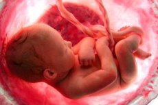
Fetal oxygen deprivation in pregnancy and labor: how to determine, what is dangerous
Last reviewed: 04.07.2025

All iLive content is medically reviewed or fact checked to ensure as much factual accuracy as possible.
We have strict sourcing guidelines and only link to reputable media sites, academic research institutions and, whenever possible, medically peer reviewed studies. Note that the numbers in parentheses ([1], [2], etc.) are clickable links to these studies.
If you feel that any of our content is inaccurate, out-of-date, or otherwise questionable, please select it and press Ctrl + Enter.
Fetal oxygen starvation is a lack of oxygen for the child in the womb. There can be many reasons for this condition, as well as consequences. It is very important to know the main risk factors for this pathology in order to control all manifestations and symptoms and correct them in time.
 [ 1 ]
[ 1 ]
Epidemiology
Statistics on the prevalence of oxygen starvation indicate that this pathology occurs in more than 40% of newborn babies. Complications of oxygen starvation can occur immediately after birth in the form of asphyxia, which occurs in 89% of babies. In the future, this can cause the death of the newborn. As for chronic hypoxia, in 30% of cases of congenital defects in a baby, it is one of the main etiological factors. In premature babies, oxygen starvation occurs very often and in 10% of children it can lead to death.
Causes fetal oxygen deprivation
The main causes of fetal oxygen starvation can be divided into several groups.
- Conditions and illnesses of the mother that affect the oxygen supply to the fetus:
- blood loss, iron deficiency anemia in the mother, which causes a lack of oxygen due to a deficiency of hemoglobin in the mother's blood;
- cardiovascular pathologies in the stage of decompensation in pregnant women;
- pathologies of the respiratory system, which are accompanied by a decrease in the level of oxygen in the tissues due to insufficient oxygen supply (hypoplasia of the lungs in the mother, tuberculosis, chronic lung diseases);
- taking medications or drugs;
- HIV infection, syphilis, chronic infectious processes of internal organs.
- Violation of intrauterine gas exchange:
- pathologies of the umbilical cord with disruption of its normal function - knots, umbilical cord entanglement;
- premature aging of the placenta;
- pathology of placenta previa;
- fetoplacental insufficiency due to illnesses in the mother;
- pathologies during childbirth that lead to prolonged standing of the fetus in the birth canal or premature detachment of the placenta.
- Conditions in the baby that may affect oxygen delivery.
- congenital defects of the nervous system (hydrocephalus, brain hernias)
- critical congenital heart or lung defects;
- intraventricular hemorrhage;
- intrauterine infections - herpes, cytomegalovirus infection, toxoplasmosis;
- Direct damage to the respiratory tract with obstruction of their patency or with serious impairment of their function.
- meconium aspiration during labor;
- tracheal hypoplasia or fusion of the airways with the esophagus, other congenital malformations of the respiratory tube.
- It should be noted that fetoplacental insufficiency is the leading cause of intrauterine hypoxia.
[ 4 ]
Risk factors
Risk factors can be identified based on the causes:
- any pathologies of pregnancy with toxicosis that can lead to disruption of placental circulation;
- extragenital diseases of the mother in a state of decompensation;
- Pathological conditions during childbirth lead to intranatal oxygen starvation.
Pathogenesis
The pathogenesis of the development of fetal oxygen starvation depends on the following factors:
- The content of oxygen and carbon dioxide in the mother's blood.
- The state of the uterine and uteroplacental circulation.
- Fetal circulation status.
Deficiency of one or more of these factors leads to a number of compensatory reactions:
- Increased placental blood flow. This helps to compensate for the lack of oxygen that occurs for a while.
- Placental hyperplasia.
- Increase in the volume of the capillary fetal zone.
- Increased fetal blood flow.
Oxygen deficiency is an ascending factor of pathogenesis. But then everything depends on the duration of oxygen deficiency. If the deficiency is temporary, then increased blood flow and other compensatory mechanisms are enough to normalize blood flow for some time. Such acute oxygen deficiency may go unnoticed by the fetus.
If oxygen deficiency is moderate but long-term, the fetus gradually adapts to it.
First of all, tissue respiration processes are intensified, anaerobic glycolysis and erythropoiesis increase, and the function of the adrenal cortex is activated. The cardiovascular system reacts by redistributing blood with preferential blood supply to vital organs - the so-called "centralization of blood circulation" (brain, heart). This slows down the capillary blood flow of parenchymatous organs. Hypoxia of muscles and internal organs leads to the accumulation of lactate and the development of metabolic acidosis.
Long-term and severe hypoxia causes a breakdown of compensation mechanisms (depletion of the adrenal cortex can lead to arterial hypotension and subsequently to shock).
Metabolic acidosis leads to increased permeability of the vascular wall, which, together with slowing blood flow and increasing concentration, leads to a sludge effect and microthrombosis. Diapedetic hemorrhages (pinpoint and large in size), cerebral edema, hypovolemia, and dysfunction of all organs and systems occur.
The most sensitive to the effects of hypoxia is the central nervous system, where the protective mechanisms of anaerobic glycolysis are most weakly expressed, therefore hypoxia develops earlier and more intensively. Disruption of ion metabolism, accumulation of toxic products causes destruction of cell components, their necrosis and death.
Pathophysiologically, two main processes develop: hemorrhagic infarction and the development of ischemia (leukomalacia).
The final result depends on the severity and duration of hypoxia, as well as the maturity of the fetus and newborn. The less mature the child, the greater the damage to the body. Traumatic factors during childbirth also play a significant role, increasing hemodynamic disorders. hypoxia, even physiological childbirth is traumatic.
Acute hypoxia, which begins during labor, differs from chronic. Due to the rapidity of this form and pathophysiological processes, the leading role here is played by immediate reflex reactions of the cardiovascular system with minimal metabolic disturbances. However, with the immaturity of the child, the influence of numerous unfavorable antenatal factors, prolonged anoxia at a certain stage, there is a breakdown of protective reactions and a sharp drop in peripheral pressure. The collapse that develops is accompanied by all pathophysiological reactions and, with chronic hypoxia, leads to hypovolemia.
Symptoms fetal oxygen deprivation
Symptoms of fetal oxygen starvation appear during pregnancy and childbirth, and the main manifestations of this are the following:
- In terms of fetal heartbeat, it increases, slows down, and then becomes arrhythmia.
- The dullness of his heart tones.
- Passage of meconium (admixture of meconium in amniotic fluid).
- Increase and then slowdown of fetal movements.
In case of diagnosis of intrauterine hypoxia, obstetricians-gynecologists must correctly determine the tactics of labor management to reduce hypoxic and traumatic damage to the child.
Primary determination of the severity of hypoxia and asphyxia of the newborn is carried out for all newborn babies according to special criteria of the Apgar scale. The assessment is carried out in the first and fifth minutes of the child's life and allows to assess the degree of adaptation of the newborn immediately after birth. This also makes it possible to suspect the presence of symptoms of oxygen starvation.
The first minute determines the severity of intrauterine hypoxic damage to the fetus.
The fifth minute determines the effectiveness of resuscitation measures and the severity of the newborn's condition. If necessary, such an assessment is carried out at the tenth and fifteenth minute.
The tenth minute determines the effectiveness of intensive therapy, depending on the adaptive mechanisms of the newborn's body.
The fifteenth minute determines the final result and prognosis of the hypoxia suffered.
Thus, the initial assessment of the child’s condition allows us to identify those children who need urgent assistance.
Considering that the disturbances during fetal oxygen starvation consist of the involvement of the brain vessels and cerebrospinal fluid in the process, a disturbance of hemo-cerebrospinal fluid dynamics occurs. This period is valid only in the first 7-10 days of the child's life. Later, if the clinical manifestations of the transferred hypoxia persist, when damage to nerve cells comes to the fore in the pathogenesis, the term "ante, intra, perinatal CNS damage of hypoxic genesis" is used. All this affects the appearance of symptoms not only in the first minutes and days of the child's life, but also throughout the month.
Nerve cells are the first to suffer in conditions of fetal oxygen starvation. Therefore, the first signs may manifest themselves as pronounced neurological disorders. Most often, this is characterized by brain hypoxia, which leads to ischemia of certain areas of the cerebral cortex. This manifests itself as a syndrome of hyperexcitability or depression of the child.
Symptoms that are characteristic of increased excitability of the child can occur already a few hours after birth. Symptoms that the mother may notice differ from those that the doctor may notice. The first signs may be in the form of frequent shrill crying of the child, the period of sleep in such children does not last more than twenty to thirty minutes. The baby's chin may shake, there may be tremors of the arms and legs when he cries. The syndrome of oppression has slightly opposite signs - the child often sleeps, his muscle tone is reduced, he lies without active movements of the arms and legs. These symptoms indicate oxygen starvation of the fetus, which develops acutely or chronically and affects the central nervous system. But with damage to the central nervous system against the background of oxygen deficiency of the fetus, other symptoms may be observed.
Convulsive syndrome can also be observed in the form of both widespread tonic-clonic seizures and local contractions of muscle groups. In this case, in newborns, the equivalent of seizures is often a spasm of the facial muscles with various facial expressions in the form of a smile, unmotivated sucking, or sticking out the tongue.
Hypertensive-hydrocephalic syndrome is accompanied by increased intracranial pressure. Clinically, this is accompanied by bulging fontanelle, divergence of cranial sutures with an increase in the volume of the child's head. Nystagmus, strabismus, and convulsive readiness may develop against this background.
The central nervous system is the main organ that suffers from fetal oxygen starvation during childbirth. After all, an acute oxygen deficiency develops, which causes such symptoms. Fetal oxygen starvation during pregnancy, which lasts for a long time, affects all the baby's organs. In this case, tissue formation processes, as well as their growth, can be disrupted. After birth, this can manifest itself in congenital malformations of internal organs. They can be from minor - in the form of a child's low body weight at birth, to significant - congenital heart defects. All this, in the absence of a direct cause, can cause complications in the child's health in the future.
Complications and consequences
What is the danger of fetal oxygen starvation? If the nervous system is affected, there may be long-term consequences in the form of residual cysts in the brain. This may not affect the child's health in any way, but there may be impairments in the child's cognitive abilities in the form of poor memory, lagging behind in school. If the damage was more serious, then impairments in the child's motor activity may develop in the form of decreased muscle tone or paralysis. Complications of oxygen starvation can be serious if this condition developed acutely during childbirth. This is accompanied by acute fetal asphyxia and the child breathes poorly and cannot establish a normal breathing rhythm and heart activity. This can even be fatal.
Diagnostics fetal oxygen deprivation
Diagnosis of fetal oxygen starvation should be as early as possible. This allows minimizing the complications that may develop against this background.
If oxygen starvation occurred during childbirth or during pregnancy, it can be diagnosed immediately after birth. In the first minute, the doctor evaluates the general condition of the baby, and if he has not closed, then the child is immediately taken to the resuscitation table and vital signs are determined - heart rate, respiratory rate, skin color, reaction to irritants. At the same time, if any of the indicators decreases, then acute asphyxia is immediately diagnosed as an external manifestation of oxygen starvation of the fetus.
But if the child was born without signs of respiratory problems, then the diagnosis of oxygen starvation can already be carried out during the examination of the child already in the ward during the first twenty-four hours after birth.
With this type of diagnosis, the doctor carefully examines the newborn in all organs and systems. The general condition of the child may be severe due to neurological symptoms - hyperesthesia, generalized clonic seizures. The color of the child's skin may be cyanotic or cyanosis may occur only in the nasolabial triangle. Reflexes: sucking, swallowing, searching, Babkin, Moro - are evoked, but they may be asymmetrical. Depending on the leading syndrome, there may be hyperreflexia or hyporeflexia. The configuration of the head may change even with a mild hydrocephalic syndrome. Also, the sagittal suture may diverge. Tissue turgor may be reduced, there may also be muscle hypotonia with oppression syndrome or pronounced hypertonia with hyperexcitability syndrome.
It is necessary to conduct diagnostics with an assessment of heart tones. Heart activity is usually rhythmic, but the tones are often weakened. All other systems are normal. Such disturbances of the general condition, tone, reflexes make it possible to assume the presence of damage to the central nervous system against the background of oxygen starvation of the fetus. In this case, additional diagnostic methods are necessary.
The tests do not give any specific changes. Instrumental diagnostics are considered more informative. All newborns with hypoxia undergo neurosonography. Neurosonography is an ultrasound method of examination (visualization) of the brain, which allows to assess the condition of the brain tissue and cerebrospinal fluid pathways through the large fontanel. Neurosonograms are used to describe the macrostructure and echogenicity of the brain tissue, the size and shape of the cerebrospinal fluid spaces, as a result of which foci of leukomalacia, intra- or periventricular hemorrhages, and expansion of the ventricular system - ventriculomegaly are detected. Hyperchoidity of the periventricular zones in the area of the anterior and posterior horns of the lateral ventricles, according to neurosonogram data, allows us to suspect periventricular leukomalacia as one of the signs of brain damage due to fetal oxygen starvation. Hyperchoidity in the subependymal areas and intraventricularly allows us to assume the presence of intraventricular hemorrhage. However, today it is believed that ultrasound diagnostics can only be used as a screening method to identify children with suspected intracranial injuries.
Characteristic changes during oxygen starvation allow us to determine that the hemispheres are symmetrical, the lumen of the lateral ventricles is not changed. This immediately excludes intraventricular hemorrhages. The detection of shadows of varying intensity and size in the brain structures, increased echogenicity of periventricular areas - all this indicates damage of hypoxic genesis. Leukomalacia in the ventricular area with subsequent formation of cysts, which can be observed throughout life, can also be observed.
Differential diagnosis
Differential diagnostics of oxygen starvation should be carried out already at the stage of clinical diagnostics. It is very important to differentiate neurological symptoms of oxygen starvation from manifestations of intraventricular hemorrhages. Intraventricular hemorrhages are usually diagnosed in premature babies born with a body weight of less than 1500 g. In contrast, oxygen starvation can occur in children of any gestational age and any weight.
The first symptoms of intraventricular hemorrhage are characterized by the clinical picture of progressive anemia, decreased muscle tone, adynamia, and bulging of the large fontanelle. Other symptoms (eye, convulsions) are less common and less pronounced (apnea attacks, tachycardia or bradycardia). Innate reflexes are depressed. Large tremors are observed, increasing with head movements, and tonic convulsions, turning into opisthotonus. Often there are disorders of the visual organ (wide open eyes, gaze paresis, flaccid pupillary response to light), vertical or rotary nystagmus, and inhibition of sucking and swallowing. That is, such symptoms, in addition to similar muscle disorders, have distinctive features - a predominance of local symptoms.
The newborn lies on its side with its head thrown back, often on this side there is a dilation of the pupil. Such meningeal symptoms are characteristic signs of hemorrhage, in contrast to the manifestations of ischemia against the background of oxygen starvation, when meningeal signs are not expressed.
Lumbar puncture reveals increased cerebrospinal fluid pressure. It is uniformly colored red or pink with a large number of fresh and altered erythrocytes.
Who to contact?
Treatment fetal oxygen deprivation
Treatment of fetal hypoxia involves providing primary care measures and treating acute damage to the nervous system.
Primary treatment for acute manifestations of hypoxia consists of measures to restore vital indicators according to the ABC system:
- Restoring air flow through the oral cavity and breathing tube (A - Airway).
- Artificial ventilation of the lungs (B - Breath).
- Indirect cardiac massage (C-Cordial).
- Correction of metabolic disorders at the end of ABC resuscitation measures.
All these treatment measures should be carried out immediately after birth and each subsequent step is carried out only if the effectiveness of the previous one has been assessed. A careful sequence of steps and feedback between them create an algorithm of the doctor's behavior in case of asphyxia. The sequence of this complex of assistance depends on the severity of hypoxia, the degree of maturity of the baby, the course of the ante- and intranatal period, as well as the effectiveness of the previous treatment, including both ante- and intranatal. The main indicators that are taken into account as control of the effectiveness of treatment measures are skin color, microcirculation disorders, hypovolemia, heart rate. The presence of one or more pathological signs determines different resuscitation tactics.
As for the treatment of oxygen starvation, which caused damage to the nervous system, a comprehensive approach is used with medications, vitamins, and restorative gymnastics.
If CNS damage is diagnosed, treatment begins with resuscitation measures in the maternity ward. Treatment of such children usually ends in the 2nd stage of premature care department or in the neonatal pathology department.
Treatment measures include:
- staying in a specialized incubator with the necessary microclimate and humidity;
- maximum protective mode (reduction of the intensity of irritants, gentle reviews, minimal pain prescriptions);
- natural feeding (depending on the severity of the condition, parenteral nutrition, feeding through a tube or from a bottle using expressed breast milk may be possible);
- carefully thought-out and appropriately limited drug therapy (dehydration, anticonvulsant, antihemorrhagic, vasoconstrictive, agents that normalize metabolic processes in the nervous tissue and increase the resistance of the brain to hypoxia).
There are no uniform approaches to prescribing certain medications. Only three clinical syndromes (hypertensive-hydrocephalic, convulsive and muscular hypertonus) reliably require prescribing medications.
It is recommended to continue breastfeeding, adhere to a daily routine, constantly stimulate visual-auditory reactions (bright toys, music, singing) and motor skills, especially motor-visual coordination during the first 3 months of life.
The main treatment objective of the syndrome of increased neuroreflex excitability is to reduce increased hypertonus and improve nerve conduction. The following approaches to the treatment of this condition exist:
- Phenobarbital is a drug from the group of neuroleptics, which acts on the central nervous system by inhibiting the activity of enzyme systems, which reduces increased nervous excitability in a child against the background of oxygen starvation. The drug also removes convulsive readiness, if the baby has a predisposition to this. The dosage of the drug is 3-4 mg / kg per day for 3-4 weeks. Side effects may occur if the dosage is violated in the form of inhibition, drowsiness, decreased reflexes, and respiratory failure. Precautions - you need to carefully combine the drug with other neuroleptics.
- Magnesium-citral mixture is a combination of 1% citral solution 2.0 milliliters, magnesium sulfate - 3.0 milliliters, valerian extract - 2.0 milliliters (or without it), 10% glucose solution - 200 ml. This combination helps reduce increased excitability of the nervous system, as well as control muscle tone and reflexes. The dosage of the drug is 1 teaspoon 3 times a day. Side effects can be in the form of depression of consciousness, allergic reactions, so for newborns the drug is used with a clear dosage and special care.
- Mydocalm is a drug used to correct increased muscle tone. It reduces the amount of active acetylcholine, which increases muscle contraction. Due to this action, pronounced hypertonicity decreases and the work of all organs and systems improves. The dosage of the drug is 0.0125-0.025 g / day. The method of application can be intramuscular, dividing the drug into two or three doses. Side effects can be in the form of twitching of individual muscle groups, severe hypotension, lethargy.
- Prozerin is a drug from the group of anticholinesterase agents. It is used in the treatment of oxygen starvation, which is accompanied by a pronounced oppression syndrome. The drug removes the action of the cholinesterase enzyme, which increases the activity of acetylcholine and improves muscle tone. The dosage of the drug is 0.003 mg / kg intramuscularly. It is used for no more than ten days to correct the tone and general condition of the child. Side effects can be in the form of convulsive contraction of facial muscles, visual impairment, allergic reactions.
- Actovegin for fetal oxygen starvation is also used in treatment to improve the recovery of damaged areas. The drug belongs to the hydrolysates of protein structure, which penetrates into the areas of ischemic areas and restores the vascular system there. This improves the recovery period. The dosage of the drug at the initial stages of treatment is up to 20 milligrams per day, divided into two doses. The method of administration is intramuscular. Then they switch to oral administration of the drug in tablets of 50 milligrams twice a day until the symptoms decrease. Side effects are disturbances in general blood flow, which may be accompanied by cold extremities.
This syndromological approach to treatment allows to correct symptoms and improve the prognosis of the child after oxygen starvation. It is very important to use vitamins and physiotherapy in the future. The most commonly used vitamins are B1, B6, ATP, 15-20 per course daily or every other day intramuscularly or by injection. Then they switch to taking oral vitamin preparations during the first month of the baby's life.
Physiotherapeutic treatment methods include general soothing massage, pine baths, paraffin applications like "boots". Electrophoresis of the cervical and lumbar spine with nicotinic acid and euphyllin is also used.
Traditional medicine is used very rarely in the acute period of fetal oxygen starvation. Such cases of using herbs or traditional medicine methods are limited to using these methods only in the late recovery period, when there are residual effects of certain symptoms.
Homeopathy can also be used to improve nerve conduction, cognitive functions. The drugs are used for a long time, sometimes during the first year of life.
Surgical treatment of this pathology is not used.
Prevention
Prevention of fetal oxygen starvation consists primarily of a safe pregnancy and bearing a healthy child. For this, any pregnancy must be planned with a thorough examination of the expectant mother. This allows eliminating risk factors on the part of the mother that can affect the disruption of oxygen delivery or the formation of the placenta.
Forecast
The prognosis for recovery in case of fetal oxygen starvation is positive, even if there were asphyxia phenomena after birth. If 15 minutes after birth the Apgar score is 0-2 points, then the mortality rate is 50%, however, in 90% of survivors normal neurological development is possible. The prognosis for complete recovery in case of hypoxic injuries is positive in case of application of all methods of treatment, massage and rehabilitation procedures in the first six months of the baby's life.
Fetal oxygen starvation is a condition in which the child, either in utero or at birth, does not receive enough oxygen for the normal development of all organs and systems. There can be many reasons for this condition and the consequences can be serious. Therefore, it is very important to prevent such conditions and promptly correct the child's condition to avoid complications.