Medical expert of the article

New publications
Fluorography in pregnancy: effect on the fetus, consequences, what is dangerous
Last reviewed: 04.07.2025

All iLive content is medically reviewed or fact checked to ensure as much factual accuracy as possible.
We have strict sourcing guidelines and only link to reputable media sites, academic research institutions and, whenever possible, medically peer reviewed studies. Note that the numbers in parentheses ([1], [2], etc.) are clickable links to these studies.
If you feel that any of our content is inaccurate, out-of-date, or otherwise questionable, please select it and press Ctrl + Enter.
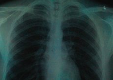
There is quite a lot of debate in the medical community about the question of whether fluorography can be done during pregnancy, because during fluorography, which is an X-ray method of visual examination of the lungs, the body is exposed to ionizing radiation. And this radiation can harm the embryo's stem cells the most.
Is fluorography mandatory during pregnancy?
However, in our reality, another question arises: is fluorography mandatory during pregnancy? And the answer to it is as follows: chest fluorography is not included in the list of tests and medical examinations that are required in women's consultations when registering pregnant women for obstetric care. At least, this is not included in the official document - Order of the Ministry of Health of Ukraine No. 417 "On the organization of outpatient obstetric and gynecological care in Ukraine" dated July 15, 2011.
But problems are still possible. When a pregnant woman first visits a women's clinic, she may be asked to fill out an Informed Consent Form, where she gives her consent, quote in translation: "to undergo all the examination methods offered to me in a timely manner (laboratory, physical, ultrasound), and, if necessary, to be examined by other specialists (if indicated)."
And further: "I confirm that the possible harm from medical interventions is less significant for me than the circumstances that prompted me to agree to them, and therefore I voluntarily and consciously give my consent to the application of the proposed set of medical interventions to me, as well as other medical interventions that will complement and ensure an adequate treatment process. However, below I indicate the medical interventions that I refuse under any circumstances, except in cases of immediate threat to my life and the life of my child or repeated agreement with me." And the medical intervention that the woman flatly refuses must be entered.
However, a woman should bring an extract from her outpatient card, which is given to the obstetrician-gynecologist by the local therapist or family doctor. It contains item 8 - Tuberculosis in the family, with subitem 8.1 - Result of fluorographic/radiological examination (indicating the date of its completion). If there is no result, then there is a reason to conduct fluorography during pregnancy...
But to detect tuberculosis – and every doctor should know this – blood is donated and an enzyme-linked immunosorbent assay (ELISA) and PCR analysis are performed; a sample of the patient’s sputum is also examined under a microscope using the Ziehl-Neelsen method, which allows the detection of Mycobacterium tuberculosis (or Koch’s bacillus).
In addition, it is useful for pregnant women to know (and for doctors not to forget) that changes in hormonal levels during pregnancy are reflected in the upper respiratory tract: swelling and hyperemia of the mucous membrane, increased secretion of mucus from the bronchi are possible, and as the gestation period increases, the chest becomes wider and the diaphragm rises several centimeters upward.
Fluorography when planning pregnancy
To make sure in advance that there are no problems with the lungs, in particular, to rule out tuberculosis, obstetricians and gynecologists advise undergoing digital fluorography when planning a pregnancy. With it, as with regular fluorography, no preparation is required.
If there is no digital fluorograph in the medical institutions of your locality, radiologists recommend undergoing a chest X-ray. Firstly, the condition of the lung tissue is much more clearly visible on the X-ray image, and it is easier for the doctor to make a diagnosis. Secondly, the single effective equivalent dose of ionizing radiation during X-ray is significantly lower than during conventional fluorography - 0.1-0.3 mSv.
It is better to plan conception at least three months after a woman has undergone fluorography.
 [
[ Technique fluoroscopy in pregnancy
Other experts claim that the technique of performing fluorography during pregnancy involves the use of a special shielding apron, which serves to protect the uterus and abdominal and pelvic organs.
There is also an opinion among doctors that any examination using X-rays during pregnancy can be carried out only for vital indications.
Vital indications during pregnancy are life-threatening situations for the expectant mother that arise in the following cases: anaphylactic reactions; pneumo- and hydrothorax (air or exudate entering the pleural cavity) and developing pulmonary atelectasis; pulmonary embolism with amniotic fluid; pulmonary thromboembolism (in pregnant women prone to deep vein thrombosis); cardiogenic shock (with hypervolemia, venous congestion in the lungs, generalized edema and tissue hypoxia); peripartum cardiomyopathy (associated with preeclampsia), etc.
But fluorography is not used in any of the above situations. You may ask, why? Because fluorography is not a diagnostic method. Western medicine, on the recommendation of the WHO, in the mid-1990s removed fluorography from the scope of diagnostic examination - due to insufficient information that the image on the fluorographic image gives the doctor. In our country and in 13 other European countries, this radiological method is used only for the primary detection (screening) of tuberculosis in the population. But even if a change in lung tissue in the form of darkening on the image is detected, a chest X-ray and appropriate tests will be required to make a diagnosis.
Contraindications to the procedure
It should be borne in mind that when performing a conventional chest fluorography (with the image saved on film), a single dose of radiation (the so-called effective equivalent dose of ionizing radiation) is 0.7-0.8 mSv (millisieverts), and the total dose per year should not exceed 1 mSv.
At the same time, some specialists believe that fluorography during early pregnancy is absolutely contraindicated, and it is permissible to conduct it only after the 20th week of gestation. In addition, it should be digital fluorography during pregnancy, that is, an examination carried out on more modern equipment. With digital fluorography, the image is recorded not on film, but on an electronic photodiode matrix, and a single dose of radiation is 0.05-0.06 mSv.
Harm of fluorography during pregnancy
How does fluorography affect pregnancy? According to research conducted under the auspices of the American Academy of Family Physicians (AAFP), the teratogenic effects of X-rays account for about 2% of all congenital intrauterine defects of the fetus.
However, there is still a lack of reliable, scientifically proven evidence of the harm that fluorography can cause to the fetus during pregnancy. Especially since the embryo (fetus) is protected in the uterus, and its radiation dose during X-ray examinations is usually lower than the dose received by a pregnant woman. And how to measure it is not yet known.
Embryonic and fetal tissues, since stem cells are in a process of constant division and differentiation, are particularly sensitive to X-rays. The consequences of fluorography – according to the concept of the non-threshold effect of ionizing radiation – can be quite serious even at low doses. Although for possible long-term consequences, exact radiation doses have not been determined, and even the time after conception (or gestational age) is approximate.
The gestational age and radiation dose are the most important factors in predicting potential effects on the fetus. The International Commission on Radiological Protection (ICRP) report, Pregnancy and Medical Radiation, noted that radiation effects on the fetus are found at approximately 50 mSv (0.05 Gy) at all stages of pregnancy. Rodent studies have shown that malformations and CNS damage may occur. It is estimated that a dose of 100 mSv (1 Gy) would likely kill 50% of embryos, and five times that dose would kill 100% of human embryos or fetuses at 18 weeks of gestation.
Experts from the US National Council on Radiation Protection and Measurements (NCRP) in the report “Radionuclide Exposure of the Embryo/Fetus” note that long-term (stochastic) effects associated with prenatal irradiation include fetal death, malformations, or an increased risk of developing cancer later in life.
Complications after the procedure
Scientific information about the effect of low doses of radiation on a fertilized egg before its implantation in the uterine cavity and in the first 3-4 weeks after conception is extremely limited. For some women who did not know about their pregnancy at the time of the X-ray examination of the lungs, the possible consequences and complications of fluorography during pregnancy of about two weeks is a miscarriage. The embryo consists of only a few cells, and damage to even one of them leads to its non-viability. But if the embryo survives, there is a high risk of gene mutations that can result in the development of congenital anomalies. Therefore, doctors recommend in such cases to undergo examination by geneticists, and at the beginning of the second trimester to undergo an ultrasound examination of the fetus.
From the third to the ninth week, the frequency of major developmental defects may increase, as at this time intensive organogenesis occurs – the laying down and formation of the fetus’s organs; growth may slow down.
Most researchers agree that during the period of 16-25 weeks of pregnancy, the dose-dependent threshold of X-ray radiation, which has a teratogenic effect (especially on brain functions), increases to 100-500 mSv (0.1-0.5 Gy), since during this period the central nervous system of the fetus is less sensitive to radiation. But this is only a theoretical assumption.
Research is underway to determine the risk of childhood cancer following prenatal exposure to ionizing radiation.