
New publications
Tartar in Dogs: Prevention and Cleaning
Last updated: 11.03.2026

All iLive content is medically reviewed or fact checked to ensure as much factual accuracy as possible.
We have strict sourcing guidelines and only link to reputable media sites, academic research institutions and, whenever possible, medically peer reviewed studies. Note that the numbers in parentheses ([1], [2], etc.) are clickable links to these studies.
If you feel that any of our content is inaccurate, out-of-date, or otherwise questionable, please select it and press Ctrl + Enter.
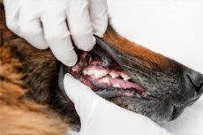
Tartar in dogs is not a disease in itself. It is mineralized plaque, a compacted bacterial biofilm that remains on the tooth surface and at the gum line for a long time. Tartar itself is important not only as a visible yellow or brown plaque, but also as a marker of chronic bacterial inflammation and a high risk of periodontal disease. [1]
The problem is extremely common. According to Cornell University, 80-90% of dogs over 3 years of age have at least one component of periodontal disease. Veterinary guidelines and surveys also indicate that periodontal disease remains the most common dental condition in dogs and often begins earlier than owners might expect based on the appearance of the teeth. [2]
The pathological process develops in stages. Initially, a soft plaque forms on the teeth, then hardens and mineralizes, turning into tartar. Bacteria at the gum line cause gingivitis, and with prolonged progression, the inflammation penetrates deeper, destroying the dental ligaments and surrounding bone, which is equivalent to periodontitis. Gingivitis is considered the only truly reversible stage, while deeper changes cause structural damage. [3]
The most frustrating aspect of this problem is that the main damage often occurs below the gum line, where the owner can't see anything. Teeth may appear relatively white in the front, but beneath the gums, pockets, bone loss, root inflammation, and pain may already be present. This is why the appearance of crowns never provides a complete picture of the oral health. [4]
It's also important to note that this isn't just a cosmetic issue or a matter of bad breath. Advanced periodontal disease is associated with pain, decreased quality of life, tooth loosening and loss, abscesses, fistulas, and, in severe cases, even pathological jaw fractures. Veterinary sources also note a link between severe oral disease and systemic changes in the heart, liver, and kidneys, although the primary clinical harm for most dogs is still localized and painful. [5]
Table 1. How the problem develops from plaque to severe periodontitis
| Stage | What's happening |
|---|---|
| Norm | The gums are pale pink, there is almost no plaque or tartar. |
| Soft dental plaque | Bacterial biofilm accumulates on the tooth |
| Tartar | The plaque mineralizes and becomes dense. |
| Gingivitis | The gums become red, swell, and may bleed. |
| Periodontitis | The ligaments of the tooth and the bone are damaged, and the tooth may become loose. |
Sources for the table. [6]
Why does tartar form and who is at risk?
The underlying trigger is always the same: plaque builds up constantly, and if not removed regularly, it becomes denser and gradually mineralizes. The rate of this process varies among dogs. In some, plaque builds up more slowly, while in others, noticeable tartar forms very quickly, which is related to the anatomy of the mouth, the microbiota, and individual characteristics of saliva and chewing behavior. [7]
Age remains one of the strongest risk factors. The older the dog, the higher the likelihood of chronic inflammation and irreversible tissue destruction around the tooth. However, don't wait until old age: many sources emphasize that a significant proportion of dogs show early signs of the disease by 2-3 years of age. [8]
Small, dwarf, and brachycephalic breeds are particularly vulnerable. They are more likely to have crowded teeth, smaller jaw volume, and unfavorable interdental geometry, which makes plaque more easily retained and reduces self-cleaning during chewing. Veterinary guidelines explicitly classify small breeds and dogs with bite anomalies as high-risk. [9]
The risk also increases with systemic conditions that impair the immune response and tissue healing. The Merck Veterinary Manual lists immunodeficiency states, diabetes mellitus, hyperadrenocorticism, poor nutritional status, and plaque-retentive anatomical surfaces, such as malocclusion, as significant factors. This is important because in such dogs, even moderate plaque accumulation can more quickly lead to complications. [10]
Finally, the dog's medical history and the quality of home care are of great importance. If a dog has had periodontitis, tooth extractions, or significant tartar in the past, it almost always requires more frequent checkups than "once a year for everyone." Current guidelines recommend personalizing the monitoring plan, as a uniform preventative schedule for all dogs is ineffective. [11]
Table 2. Main risk factors
| Factor | Why is the risk higher? |
|---|---|
| Age over 3 years | The disease accumulates with age |
| Small and dwarf breeds | Crowding of teeth is more common |
| Brachycephalic breeds | The anatomy of the skull impairs self-cleaning |
| Malocclusion | The raid is easier to delay |
| Diabetes mellitus and other systemic diseases | Poorer inflammation control and healing |
| Previously suffered periodontitis | Higher risk of recurrence |
| Irregular home hygiene | Plaque mineralizes faster |
Sources for the table. [12]
How to recognize tartar and understand that the problem is already serious
The most common early sign is persistent bad breath. Many owners dismiss it as "normal dog odor," but veterinary sources emphasize that a pronounced bad odor is often one of the first external indications of periodontal disease. It appears long before the dog stops eating or becomes noticeably sore. [13]
The next typical sign is visible yellow, brown, or grayish deposits at the gum line. These are often accompanied by redness, swelling, and bleeding of the gums. However, it's important to remember that the amount of visible tartar doesn't always accurately reflect the depth of the problem, as a significant portion of the lesion is located subgingivally. [14]
As the pain and inflammation intensify, the dog's behavior changes. It may chew on one side, be cautious when grasping toys, drop food, swallow with discomfort, drool profusely, shake its head, click its jaw, or rub its face with its paw. Some dogs show very few signs, even with severe disease, so the absence of obvious complaints does not rule out a serious lesion. [15]
If the process has progressed, more severe symptoms appear: tooth mobility, refusal to eat hard food, blood in the saliva, pain when touching the muzzle, swelling under the eye or on the muzzle, a fistula, and pronounced apathy. Such signs suggest not only tartar, but also a root abscess, deep periodontitis, a fractured crown, or another serious dental condition. [16]
It's especially important to understand that white, visible surfaces of teeth do not guarantee health. After superficial tartar removal without a full veterinary examination, the mouth may appear "cleaner," but the disease beneath the gums may still persist. This false visual "normality" is considered one of the reasons for late diagnosis in dogs. [17]
Table 3. Symptoms and their meaning
| Sign | What could it mean? | Urgency |
|---|---|---|
| Bad breath | Early sign of plaque, gingivitis, periodontitis | Scheduled inspection |
| Visible yellow or brown stone | Mineralized plaque and chronic inflammation | Scheduled inspection |
| Red and bleeding gums | Gingivitis or a more serious lesion | Inspection coming soon |
| Chewing on one side, food falling out | Pain, tooth mobility, fracture, deep periodontitis | Inspection coming soon |
| Saliva with blood | Active inflammation, trauma, gum disease | Inspection coming soon |
| Swelling under the eye, fistula, sharp pain | A tooth root abscess is possible. | Urgent visit |
| Loose tooth | Marked loss of supporting tissues | Urgent visit |
Sources for the table. [18]
Diagnosis by a veterinarian
An initial awake examination is useful, but its capabilities are limited. While the dentist can see calculus, gum inflammation, obvious crown fractures, and some tumor-like lesions, a full assessment of periodontal pockets, roots, and bone condition is impossible without general anesthesia. This is a key point that is often overlooked by owners. [19]
Current veterinary guidelines consider a full dental examination to be part of the procedure under general anesthesia. After intubation and patient stabilization, a full examination of the entire oral cavity is performed, including probing, mapping of lesions, and assessment of all dental surfaces. The World Small Animal Veterinary Association specifically states that all dental procedures should be performed under general anesthesia. [20]
Dental radiography is especially important. American Veterinary Guidelines strongly recommend full-mouth intraoral radiographs for all dental patients because without them, it is impossible to fully assess roots, bone, hidden resorption, the depth of tissue loss, and the vitality of individual teeth. In other words, without radiography, it's easy to miss exactly what's causing the most pain. [21]
Based on the examination, the doctor determines the stage of the disease. The American Veterinary Medical Association uses a practical scale from 0 to 4, where stage 1 corresponds to early gum inflammation with visible tartar, stages 2 and 3 already include bone loss visible on radiographs, and stage 4 signifies severe destruction with gum recession, significant calculus accumulation, and significant loss of supporting tissue. [22]
A diagnosis isn't just about confirming the presence of tartar. It determines the entire treatment plan: whether the tooth can be saved, whether extraction is necessary, whether there is an abscess, how aggressive the treatment should be, what pain control is needed, and how often the dog should be seen for follow-up examinations. That's why high-quality canine dentistry always begins with a thorough assessment, not simply removing visible plaque. [23]
Table 4. What is included in a complete veterinary dental examination?
| Stage | What is it for? |
|---|---|
| Conscious examination | Initial assessment of visible problems |
| General anesthesia and intubation | Safety and the possibility of a complete examination |
| Examination of the entire oral cavity | Detection of hidden lesions of soft tissues and teeth |
| Probing and mapping | Assessment of pockets, mobility, recession |
| Full-mouth radiography | Checking the roots and bone under the gum |
| Diagnostic classification of stage | Choice of treatment and prognosis |
Sources for the table. [24]
Table 5. Practical scale of periodontal disease severity
| Stage | A typical picture |
|---|---|
| 0 | Normal, no pronounced inflammation |
| 1 | Visible tartar, redness and slight swelling of the gums |
| 2 | More visible inflammation and initial bone loss on radiography |
| 3 | More severe bone loss and deep involvement of supporting tissues |
| 4 | Heavy tartar, gum recession, severe destruction and high risk of tooth loss |
Sources for the table. [25]
Treatment and professional cleaning
Professional dental cleaning under general anesthesia remains the standard of care. According to veterinary guidelines, this cleaning includes removal of supragingival and subgingival calculus, polishing of teeth, and a full dental examination performed by a trained professional under general anesthesia. This differs fundamentally from superficial cosmetic scaling. [26]
During the procedure, plaque and tartar are removed from both the supragingival and subgingival surfaces, as subgingival deposits are the most significant contributor to the progression of periodontitis. After plaque removal, the teeth are polished, as scaling creates a micro-rough surface, and without polishing, plaque will adhere more quickly. If necessary, topical treatment, extraction of non-vital teeth, and other interventions are performed. [27]
If the disease is advanced, cleaning alone won't solve the problem. Severe periodontitis, significant bone loss, tooth mobility, fistulas, abscesses, or non-viable roots may require tooth extraction. More complex methods of preserving individual teeth are sometimes available in specialized dentistry, but in general practice, the most common solution for a hopeless tooth remains extraction as a way to remove the source of chronic pain and infection. [28]
Owners are often wary of general anesthesia and therefore seek "anesthesia-free cleaning." However, the American Animal Hospital Association, the World Small Animal Veterinary Association, and the American Veterinary Dental College all consider anesthesia-free cleaning inappropriate. The reasons are the same: stress and pain for the animal, risk of aspiration and injury, inability to reach subgingival tissue, inability to perform a full examination and radiographs, and a false sense of well-being after temporary whitening of the visible surface of the teeth. [29]
Modern veterinary anesthesia is not simply a matter of "injection and cleaning." Professional guidelines describe preliminary patient assessment, vital signs monitoring, pain management, warming, intubation, and wake-up monitoring. This doesn't eliminate risks entirely, but it does manage them and makes the benefits of a high-quality dental procedure significantly outweigh the risks associated with properly managed procedures. [30]
It's also crucial to understand that even the most ideal home cleaning is no substitute for professional sanitation if your dog already has tartar and inflammation. American guidelines clearly state that brushing removes plaque, but does not remove established tartar, and daily home hygiene does not replace the need for anesthetic examinations, X-rays, and dental treatment. Home care is needed not instead of professional cleaning, but after and between procedures. [31]
Early follow-up after the procedure is beneficial. The American Animal Hospital Association recommends scheduling a follow-up visit 10-14 days after even limited preventative debridement to assess healing, discuss home care, and adjust the long-term plan. This is especially important for dogs with previous extractions, deep pockets, or poor owner compliance with home care. [32]
Table 6. What really helps and what creates a false sense of security
| Approach | Grade |
|---|---|
| Professional sanitation under general anesthesia | The main and correct method of treatment |
| Full-mouth radiography | It is necessary to search for hidden pathology. |
| Subgingival scaling | Essential for disease control |
| Polishing after stone removal | Needed to reduce plaque re-adhesion |
| Home brush after sanitation | The main method of preventing relapse |
| Chewables, special diets, and supplements with proven effectiveness | Useful as a supplement |
| Superficial "cleaning without anesthesia" | Does not treat subgingival disease and is not recommended |
| Random rare house cleaning | Insufficient for reliable prevention |
Sources for the table. [33]
Home prevention and long-term care
The best method of prevention is regular toothbrushing. Current veterinary recommendations call toothbrushing the most effective method of plaque control at home. The World Small Animal Veterinary Association recommends making brushing a daily habit, and the American Veterinary Medical Association notes that daily brushing is optimal, although several times a week is also better than no brushing. [34]
It's best to begin training gradually. The World Small Animal Veterinary Association recommends simply touching the face and lips first, then touching the teeth with your finger, then gently massaging the gum line, and only then moving on to brushing. This step-by-step approach reduces resistance and helps turn the procedure into a routine rather than a daily struggle. [35]
For brushing, use only veterinary toothpaste designed for oral use. Human toothpastes are not suitable for dogs: veterinary sources warn of problems with swallowing regular toothpaste, and xylitol is toxic to dogs. The brush should be soft, and focus on the gum line and the outer surfaces of large teeth, where plaque and tartar accumulate most quickly. [36]
Additional products may be helpful, but only as a supplement to brushing. The Veterinary Oral Health Council maintains a list of products that have been shown to slow plaque and/or tartar accumulation, and veterinary guidelines recommend focusing on these proven products. A separate 2025 study found that mechanical methods, such as brushing and daily chews, do reduce plaque buildup, but do not replace professional scaling when indicated. [37]
A dog still needs regular checkups with a veterinarian, even if the owner brushes their teeth perfectly at home. The World Small Animal Veterinary Association recommends regular dental checkups, at least annually, and more frequent checkups are required for dogs at risk. The long-term prognosis is usually good if the disease is not neglected: with high-quality sanitation and proper home care, pain, odor, inflammation, and the risk of tooth loss can be reduced for a long time. [38]
Table 7. Practical home care plan
| Step | How to do it right |
|---|---|
| Training | Start with touching the lips and teeth, without forcing |
| Frequency | Strive for daily cleaning |
| Paste | Use only veterinary |
| Brush | Soft, properly sized mouth |
| Cleaning area | First of all, the gum line and the outer surfaces of the teeth |
| Additions | Use only proven aids |
| Inspections | At least once a year, in dogs the risk is higher |
| After rehabilitation | Follow an individual plan for follow-up visits |
Sources for the table. [39]
Frequently asked questions
1. Is tartar in dogs simply a cosmetic problem?
No. Tartar is important as a visible sign of chronic bacterial inflammation. The main damage is not related to tooth color, but to gingivitis, periodontitis, pain, and tissue destruction under the gums. [40]
2. If the teeth look light, does that mean everything is fine?
Not necessarily. Much dental pathology in dogs is located below the gum line and may not be visible without an anesthetized examination and x-rays. [41]
3. Is it possible to remove tartar at home with a brush?
A brush is effective at removing soft plaque and slowing the formation of new tartar, but it doesn't remove hardened tartar that has already formed. Professional treatment is usually required for this. [42]
4. Why isn't cleaning without anesthesia considered a proper treatment?
Because it doesn't allow for subgingival treatment, doesn't allow for a full examination and x-ray, can be painful, and creates the false impression that the problem has been resolved. [43]
5. How often should you brush your dog's teeth at home?
The optimal goal is daily brushing. If daily brushing isn't possible, a few times a week is still better than no brushing, but it's less effective than a daily routine. [44]
6. Are human toothpastes suitable for dogs?
No. Only veterinary toothpastes that are safe to swallow are needed. Xylitol is toxic to dogs, and regular human toothpastes are not designed for ingestion. [45]
7. Can chew treats replace brushing?
No. They can be a useful addition, especially if they have been proven effective, but are not considered a complete replacement for regular brushing and professional examinations. [46]
8. When should you see a doctor urgently, rather than wait for a scheduled appointment?
You should make an appointment quickly if your pet's face is swollen, there's a hole under the eye, blood in the saliva, refuses to eat, there's severe pain, a loose tooth, or a sharp deterioration in overall health. These symptoms may indicate not just a simple calculus, but an abscess, deep periodontal disease, or another serious condition. [47]
9. Is it true that small dogs are more likely to suffer?
Yes. Small, dwarf, and brachycephalic breeds are at increased risk due to their closely spaced teeth and jaw structure. [48]
10. Is it possible to forget about the problem for a long time after one professional cleaning?
No. Professional cleaning removes existing deposits and treats the current pathology, but plaque begins to form again, so without home care and follow-up examinations, the disease returns. [49]