Medical expert of the article

New publications
Kephalohematoma in newborns on the head: causes, how to treat, prevention
Last reviewed: 04.07.2025

All iLive content is medically reviewed or fact checked to ensure as much factual accuracy as possible.
We have strict sourcing guidelines and only link to reputable media sites, academic research institutions and, whenever possible, medically peer reviewed studies. Note that the numbers in parentheses ([1], [2], etc.) are clickable links to these studies.
If you feel that any of our content is inaccurate, out-of-date, or otherwise questionable, please select it and press Ctrl + Enter.
Cephalohematoma in a newborn is a blood accumulation on the head within the bone, which occurs in a child due to the peculiarities of the birth process itself. This is a very common pathology today due to the increase in the number of complicated births. A feature of cephalohematoma is an individual approach to treatment, which every mother should know.
Epidemiology
Statistics of births of children with cephalohematomas show that one out of 200 newborns has this problem. In 45% of cases, cephalohematoma is combined with a birth tumor. In healthy full-term children with cephalohematoma, more than 67% of cases of pathological births. As for premature babies, about 15% of such newborns have cephalohematoma, regardless of the course of the birth itself.
Causes neonatal kephalohematoma.
To understand the causes of cephalohematoma, you need to know what this concept means. It is a collection of blood, which thickens over time, within one bone under the periosteum. It is very important that the blood accumulates within the bone and does not spread further.
What are the causes of cephalhematoma on the head of a newborn? The main cause of any hematoma is external influence in the form of a blow or physical impact. Therefore, the cause of cephalhematoma formation is most often complicated childbirth. But here we are not necessarily talking about the intervention of doctors during childbirth, but rather the opposite - the lack of active tactics of childbirth can lead not only to the formation of cephalhematoma, but also to other complications.
The cause of cephalohematoma formation can be considered a pathology of the pelvic structure, which can be caused by injuries or incorrect turns of the child. Hematomas are often observed in the case of using obstetric forceps or simple extraction of the fetus.
Even without the absence of an obvious cause, cephalohematomas can be observed due to simple weakness of the vascular wall. Most often, such pathology occurs in premature babies. This is due to the fact that the bone structure has its own characteristics - the germinal matrix is highly developed, which is very easily injured. Therefore, even with normal birth, a cephalohematoma can form in a premature baby.
Risk factors
Based on these reasons, it is necessary to identify risk factors for the development of cephalohematoma. These include pathological pregnancy, complicated labor, interventions during labor, and the birth of a premature baby. All of this is a potential threat to the development of such a pathology.
Pathogenesis
The pathogenesis of cephalohematoma formation is the rupture of blood vessels that supply the bone, and blood can freely flow out under the periosteum. This is accompanied by the formation of a hematoma within the bone where the rupture of the vessels occurred. Such a rupture of vessels can occur with prolonged compression of the tissues of the head, for example, with weak labor activity, when the baby's head is in one plane of the pelvis for a long time. This disrupts the normal outflow of blood and with further movement of the fetus, hemorrhage under the periosteum can occur.
 [ 12 ]
Symptoms neonatal kephalohematoma.
The first signs of cephalohematoma appear after birth, and they are very objective. Even the mother can see the asymmetry of the child's head due to a tumor of different sizes. Such asymmetry can be different and depends on the location of the hemorrhage. Most often, you can see a cephalohematoma of the left or right parietal region of a newborn, sometimes the occipital region, since these places are most susceptible to compression at birth. Symptoms of cephalohematoma are characterized by the presence of a tumor of clear dimensions corresponding to the size of the bone. Such a tumor slightly fluctuates when pressed, can be blue. It does not affect the general condition of the child and can be easily tolerated by the child. The only unpleasant symptom can be considered asymmetry of the child's head.
Stages
The stages of development of cephalhematoma do not differ from those of hematomas located elsewhere. Naturally, the duration of each stage depends on the amount of blood in the tissues. At the beginning of development, cephalhematoma is an accumulation of liquid blood that has spilled into the surrounding tissues. Then, after a few hours, the blood begins to gradually thicken. Then there will be no more "fluctuation" symptom. Over time, such blood gradually dissolves and the hematoma passes. How does cephalhematoma resolve in newborns? The process begins from the center in such a way that a small ridge of blood is formed along the edges, which has not yet been completely lysed. In this case, the blood breaks down into its components and bilirubin is formed, which can cause transient jaundice in the child. Therefore, one of the symptoms of cephalhematoma in a newborn can be the appearance of jaundice at the time of its resorption. As a rule, it should not exceed physiological limits, but if the cephalohematoma is very large, then the child’s condition must be closely monitored.
[ 15 ]
Complications and consequences
Is cephalohematoma dangerous in newborns? It is difficult to say for sure. But the symptoms of a small cephalohematoma do not cause any harm to the child, do not disrupt the child's nutrition, do not hurt, and the body temperature does not rise. But if the hematomas are of significant size, then there is already a risk of complications. The most frequent complications can develop with significant hematomas, when the volume of blood is impressive. In this case, the newborn may develop anemia with subsequent severe jaundice.
One of the consequences can also be considered ossification of cephalhematoma in newborns. This can occur when not all the blood is completely absorbed and a bone ridge is formed along the edges. It can be small in size and may not cause any concerns. The only thing is that there may be a cosmetic defect in boys, and even then it often goes away with the growth of the skull bones. One of the frequent complications can also be infection with the development of septic conditions. This can happen with surgical intervention in the form of a puncture. Therefore, the consequences and complications of cephalhematoma directly depend on its size and treatment tactics.
[ 16 ]
Diagnostics neonatal kephalohematoma.
Diagnosis of cephalohematoma in newborns is not difficult, since visually it is a very clear picture and it is one of the few diagnoses that can be seen. The main role here belongs to differential diagnostics. But despite this, with large sizes of cephalohematoma there is a need to monitor the child's condition. In case of severe jaundice, it is necessary to conduct tests to determine the level of bilirubin in the blood.
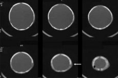
Instrumental diagnostics should be performed to exclude complications and clarify the diagnosis. Ultrasound diagnostics allows you to determine the location of the hematoma, as well as accurately determine the amount of blood. In dynamics, you can watch how the resorption occurs and the need for active treatment of the pathology.
If the baby is premature and has a cephalohematoma, it is also recommended to perform an ultrasound of the brain to assess the state of the nervous system.
[ 17 ]
What do need to examine?
Differential diagnosis
Cephalhematoma is an extracranial hemorrhage, so it is necessary to conduct differential diagnostics with other similar pathologies - subgaleal hematoma and birth tumor.
Subaponeurotic hematoma is a blood accumulation in the space between the periosteum and the aponeurosis of the tendon helmet. Such a tumor has huge dimensions and is located from the eyebrows to the back of the head. And the main differential feature of cephalohematoma is its location within one bone.
A birth tumor is a soft tissue swelling of the skull without hemorrhage. This occurs when the fetus stands in one plane for a long time, which disrupts the outflow of venous blood from the head and causes such swelling. Therefore, a distinctive feature of a birth tumor is its correspondence to the presentation of the child. It passes faster than a cephalohematoma and there is no fluctuation.
Who to contact?
Treatment neonatal kephalohematoma.
Approaches to the treatment of cephalohematoma are very different and today there is no single tactic for treating the pathology. Different clinics have different experience and their own approach to this problem, so the treatment may differ. And it is impossible to say that one tactic is better than another, since different cases differ from each other.
Conservative treatment of cephalohematoma involves only monitoring the tumor regardless of its size. This is explained by the fact that additional external intervention always increases the risk of external infection. Therefore, some doctors monitor the hematoma until it is completely absorbed. In this case, the child's condition, tests, degree of jaundice and other manifestations are monitored.
There is a treatment tactic, in which only a small hematoma is subject to observation, and if its size is significant, then the rate of its spontaneous resorption is very low. Therefore, surgical treatment is offered. In this case, puncture of cephalhematoma in a newborn is used most often. This allows you to suck out a larger amount of blood, and what remains can resolve itself. In this case, all conditions should be used to minimize external infection.
Removal of cephalohematoma in newborns can be used when there is a large accumulation of blood that has already partially coagulated and cannot resolve on its own. Sometimes there is a very large clot that cannot be cured in any way except for active surgical intervention.
Medicines for the treatment of cephalhematoma do not have an evidence base for their effectiveness. It can be said that the child's body itself can cope with a cephalhematoma of any size better and faster than external factors. However, various ointments, creams, and gels are used to improve blood circulation and more active resorption of the hematoma. Troxevasin for cephalhematoma in newborns is used quite often as a topical agent. It improves local blood circulation and lymph drainage, which accelerates the process of resorption of tumor remnants.
But in any case, when choosing a treatment strategy for cephalohematoma, the mother should consult with the doctor. It is necessary to clarify all the treatment options that the doctor offers in this case and talk about the possible consequences. The parents decide anyway, but they should know about all the nuances and possibilities of treating such a condition in a child.
Prevention
Prevention of cephalohematoma is a difficult task, since it is very difficult to predict such a pathology. But given that it occurs in mothers with complicated pregnancy and childbirth, as well as in premature babies, the main method of prevention can be considered a normal pregnancy. Mom and dad should take care of their health, remembering the health of their future children.
Forecast
The prognosis for a favorable resolution of cephalohematoma is positive, since the pathology does not often have complications.
A cephalohematoma in a newborn is not necessarily associated with an incorrectly performed birth by a doctor. This condition can also develop in a healthy baby, so there is no need to panic. Expectant tactics with minimal external interventions are considered the most acceptable method of treating cephalohematomas.