Medical expert of the article

New publications
Hematoma in pregnancy
Last reviewed: 05.07.2025

All iLive content is medically reviewed or fact checked to ensure as much factual accuracy as possible.
We have strict sourcing guidelines and only link to reputable media sites, academic research institutions and, whenever possible, medically peer reviewed studies. Note that the numbers in parentheses ([1], [2], etc.) are clickable links to these studies.
If you feel that any of our content is inaccurate, out-of-date, or otherwise questionable, please select it and press Ctrl + Enter.
It may happen that the expectant mother feels fine, nothing bothers her, but when she comes to the ultrasound, she finds out that she has been diagnosed with a hematoma during pregnancy. An ordinary person understands that a hematoma is a hemorrhage into the tissue layer, which occurs as a result of an injury.
Today, doctors also name other causes of this pathology.
 [ 1 ]
[ 1 ]
Causes pregnancy bruises
Modern obstetricians and gynecologists are increasingly faced with the problem of carrying a fetus by a woman who has been diagnosed with intrauterine hemorrhage. Quite often, it develops against the background of fragile blood vessels that supply nutrition to the uterus and placenta.
The following are the main causes of hematoma during pregnancy:
- A disruption in a woman's metabolic processes.
- Inflammatory and infectious diseases affecting the genitourinary system of the expectant mother.
- A severe degree of toxicosis, which is especially dangerous in the late stages of pregnancy.
- Sharp jumps in blood pressure. High numbers are especially dangerous.
- Strong stressful situations.
- The cause of a hematoma during pregnancy may also be a pathology in the development of the fertilized egg.
- Hormonal imbalance.
- Problems observed during blood clotting, pathological changes in the walls of the vessels of the placenta and the entire body of the woman.
- Abuse of alcohol and smoking during the period when a woman is carrying a child.
- Congenital or acquired pathology of the development of the uterus.
Symptoms pregnancy bruises
Waiting for the birth of a child is a state of complete happiness, a time of hope and one of the most enchanting moments in the life of any woman. But at the same time, it is anxiety and worry about the health of the unborn baby. A hematoma in the uterus during pregnancy is a difficult test for a woman expecting a child. After all, in its presence, the fertilized egg begins to peel off from the surface of the uterine endometrium, blood gradually accumulates in this place, forming a bruise.
A hematoma in the uterus during pregnancy poses a serious danger to bearing a child, as it can provoke a spontaneous miscarriage, especially in severe forms of the pathology. In mild cases, a woman may not even notice it and learn about its existence after a successful resolution of childbirth.
A severe degree of hematoma development during pregnancy is dangerous not only for the baby, but also for its mother. Bleeding provokes a deterioration in the mother's general health, and posthemorrhagic anemia develops. Due to a failure in blood circulation, the fetus receives less oxygen, which leads to "oxygen starvation", and this, in turn, to a slowdown in the child's development, both physically and psychologically.
Hematoma in early pregnancy
For many, the word "hematoma" is directly associated with a serious injury. Therefore, having heard such a diagnosis and not fully understanding its essence, a pregnant woman begins to get nervous. And it is understandable. After all, a hematoma in the early stages of pregnancy can indicate the beginning of the rejection of the fertilized egg, which leads to a miscarriage (termination of pregnancy). By the way, this pathology is not very rare, and the highest percentage of its manifestation occurs in the fifth to eighth week of pregnancy.
There are many reasons for this pathology: hormonal imbalance, a burdened hereditary history, sexual intercourse, great nervous or physical overexertion.
But diagnosing a hematoma in the early stages of pregnancy allows you to undergo a course of treatment as early as possible, which will ensure normal development of the fetus and a successful birth.
Quite often, the expectant mother does not even suspect the existence of an internal hematoma, and for her, the most complete surprise is its presence, which was discovered during a routine ultrasound examination. Symptoms of a hematoma during pregnancy mainly depend on the severity and localization of the pathology.
- Mild severity. In this case, the pregnant woman feels normal, no physical manifestations of internal hemorrhage are determined. Hematoma during pregnancy is diagnosed only by ultrasound or after the birth has successfully ended naturally, since it leaves its marks on the placenta.
- Moderate severity. In this case, the woman experiences pulling, aching pain in the lower abdomen. Red or brownish discharge may appear from the genital tract. If such symptoms are present, you should immediately seek help from an obstetrician-gynecologist who is monitoring the pregnancy. Whether or not there is discharge is largely determined by the volume of the hematoma and its location. In the case of moderate pathology, an irregular heartbeat is heard in the unborn baby.
- Hematoma during pregnancy of severe severity. The intensity of blood discharge increases, and the pain in the lower abdomen increases. It becomes cramping. Against the background of the bleeding that has opened, the expectant mother's blood pressure drops. The woman may even lose consciousness.
Any practicing physician knows that if a diagnosis of retrochorial hematoma is made, this means that there is a direct threat of termination of pregnancy.
Discharge from hematoma during pregnancy
If during the first trimester of pregnancy a woman observes light brown vaginal discharge, but does not feel any discomfort or deterioration in health, there is no need to be particularly concerned, even if an ultrasound examination (which every pregnant woman undergoes routinely) reveals a hematoma during pregnancy.
Another question is if the color of the discharge is bright red. This already indicates that the bleeding does not stop. In this situation, it is necessary to urgently take emergency measures, otherwise premature rejection of the fetus may occur, and the woman will lose the child.
[ 7 ]
Forms
We are used to seeing various types of hemorrhages on the skin surface, but many did not even guess that there are also uterine hematomas that occur on the uterus during the period when a woman is carrying her baby. There is a classification of this pathology. The following types of hematomas are differentiated during pregnancy:
- Clinic and period of development of the disease:
- Retrochorial. This type of effusion is diagnosed in the early stages of pregnancy (up to 16 weeks) and is a detachment of the fertilized egg from the chorion (the outer embryonic membrane).
- Retroplacental. By the 16th week of pregnancy, with normal fetal development, the placenta is already fully formed. If a hematoma develops later, it provokes placental abruption before the due date, which is fraught with spontaneous termination of pregnancy, i.e. the woman loses her unborn child.
- By severity of pathology:
- Mild pathology.
- Moderate degree of disease manifestation.
- Acute, severe hematoma during pregnancy.
A mild or moderate hematoma during pregnancy is an indication for a cesarean section, and the operation is scheduled for an earlier period than the time set for natural childbirth.
Retrochorial hematoma in pregnancy
The chorion is the outer embryonic membrane that surrounds the embryo and is formed in the early stages of the gestational period, being the predecessor of the placenta. This is where the retrochorial hematoma during pregnancy gets its name, which is due to its timing of origin and location. This pathology develops as a result of the detachment of the ovum from the chorion. It is observed in the first trimester of pregnancy, before the transition to the second. Blood gradually begins to accumulate at the site of rejection - a hematoma is formed, a clear sign of the threat of spontaneous abortion.
This pathology is accompanied by vaginal discharge of a slightly brownish hue. At the same time, the woman experiences a nagging pain in the lower abdomen. If the hematoma is localized at the bottom of the uterus, there may be no obvious symptoms of the pathology. Then the hemorrhage can only be detected using ultrasound.
When brown discharge appears, a woman begins to worry about the fate of her pregnancy, obstetricians and gynecologists do not consider this a bad symptom. Since the blood accumulates in the cavity between the membranes of the chorion and the fetus, coagulates there, taking on a brownish tint, discharge of this color, on the contrary, may indicate that blood clots are gradually beginning to come out, "resolving" the hematoma.
When it is really necessary to sound the alarm, it is when the discharge is scarlet. This is a clear sign that the bleeding has not stopped, the hematoma continues to grow, the fertilized egg continues to peel off and if emergency measures are not taken, the woman in labor may lose the child, since in such a situation a premature termination of pregnancy occurs. In this case, in order to assess the growth of the hematoma during pregnancy, the doctor prescribes not only an ultrasound, but is also obliged to monitor the level of fibrinogen in the blood plasma.
Retroplacental hematoma in pregnancy
In the case where the rejection of the fertilized egg occurs from the chorion, as a rule, this occurs in the first trimester, a retrochorial hematoma develops. If this process begins later (after 22 weeks of pregnancy), when the chorion has degenerated into the placenta, the same process of rejection of the embryo is called a retroplacental hematoma. It occurs according to the same scenario as in the first case, when the appearance of bleeding is a real threat of termination of pregnancy.
Similarly, retroplacental hematoma during pregnancy is the main symptom indicating a risk of miscarriage. The clinical picture of this pathology is: a nagging pain in the lower abdomen, bloody discharge, increased uterine tone... The behavior of the fetus itself changes: its motor activity increases, negative changes occur in the heart rhythm (tachycardia is initially heard, and then bradycardia), which indicates a violation of its normal development. The obstetrician-gynecologist interprets these signs as untimely rejection of placental tissue, which can lead to a miscarriage.
In this case, an ultrasound scan helps in early diagnosis of retroplacental hematoma during pregnancy, which allows timely and adequate measures to save the fetus and the mother herself.
Subchorionic hematoma during pregnancy
Subchorionic hematoma is most common during pregnancy. This is a rather dangerous type of hemorrhage. It requires immediate attention from your doctor. In case of differentiation of this pathology, the size of the hematoma must be observed dynamically.
Retroamniotic hematoma in pregnancy
Retroamniotic hematoma during pregnancy is diagnosed already at the first ultrasound examination (approximately 12 weeks of pregnancy), often there is no blood discharge, but still, to be on the safe side, the obstetrician-gynecologist monitoring the pregnancy can admit the expectant mother to the obstetrics and gynecology department in order to observe the growth of the hematoma in the process.
Subamniotic hematoma during pregnancy
It does not pose a danger to the normal development of the future child. In the future, the hematoma, especially if its location is the cervical os, can dissolve on its own or come out in the form of clots.
Intrauterine hematoma during pregnancy
This is a fairly serious pathology. External or internal causes lead to the detachment of the fertilized egg, which provokes bleeding, and then a hematoma forms at the site of rejection. If the area of the hemorrhage does not increase, and the pregnant woman feels satisfactory, the embryo develops normally - then there should be no reason to worry. If the obstetrician-gynecologist sees the progression of the pathology, the question of hospitalization of the woman in labor and her treatment is raised.
Complications and consequences
The birth of a new person is the greatest mystery that nature has given to man. But how much the expectant mother has to worry about before the moment when her baby is born. Hemorrhages of various localizations. How dangerous are they? What are the consequences of a hematoma during pregnancy? Naturally, complications can manifest themselves, or everything can go well. Much in this problem depends on the parameters of the hematoma. The most dangerous is a hematoma that occupies at least 40% of the total area of the embryonic membrane and the volume of which exceeds 20 ml. Such a hematoma can slow down the growth and full development of the fetus, and cause spontaneous termination of pregnancy. Inhibition in the growth of the CTE (coccygeal-parietal size) for more than ten days indicates an increased risk of an unfavorable outcome of bearing a child.
The most dangerous consequence of such a course of events may be "Kuveler's uterus". Premature detachment of the placenta leads to the endometrium filling with blood, foci of necrosis appearing in it, which is already an indication for a complete resection of the uterus. And this is already a death sentence - a woman will never be able to become a mother.
Also, with a hematoma during pregnancy, there is a high probability of heavy bleeding, the consequences of which are quite difficult to predict, since it can even lead to a fatal outcome, both for the child and for the mother herself.
But if a pregnant woman has undergone effective treatment in a timely manner, the chances of giving birth to a normally developed baby on her own and on time increase dramatically.
Why is a hematoma dangerous during pregnancy?
The first question a woman asks after (after undergoing an ultrasound) she has heard this diagnosis is: "What is the danger of a hematoma during pregnancy?" The answer to this question largely depends on the size of the hematoma, the time of its manifestation and its localization. If a hematoma is diagnosed in the first trimester of pregnancy, this is fraught with spontaneous termination of pregnancy. If the pathology occurs at a later stage, the consequences of its appearance can affect the development of the fetus (delay in physical development, hypoxia (insufficient amount of oxygen for the full development of the child)).
The most dangerous in this category are effusions of 20 ml or more, which is about 40% of the volume of the ovum. In addition to the inhibition of the development of the fetus itself, the risk of further placental abruption increases. If the CTE (crown-rump length) of the embryo is delayed in growth by more than ten days, this is a bad sign, which indicates a high probability of an unfavorable outcome of pregnancy. Although with adequate therapy the fetus continues to develop normally, but in this case a cesarean section is indicated.
Diagnostics pregnancy bruises
It is advisable to diagnose any pathology at the early stages of its manifestation, when there have not yet been any cardinal negative changes that cannot be corrected. Therefore, in order for the pregnancy to end with normal childbirth, hematoma diagnostics are performed during pregnancy.
The main source of information during this period is ultrasound examination (US) - this is a modern, fairly informative, method of examination. In addition, the obstetrician-gynecologist prescribes other examinations.
- Complete clinical blood test
- General urine analysis.
- Blood tests for RW and HIV are performed.
- Coagulogram. Determination of the prothrombin index (PTI), which shows how well the blood clots, as well as the activated partial thromboplastin time (APTT).
- A smear of microflora taken from the vagina.
- Biochemical blood test.
- Screening for various sexually transmitted infections (STIs).
- Dopplerometry (one of the types of ultrasound, used to assess the nature and speed of blood flow in the vessels, in this case in the baby and in the placenta).
- If necessary, a blood test for hormones is prescribed.
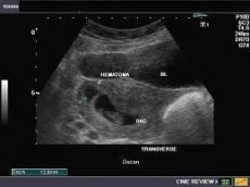
Hematoma during pregnancy on ultrasound
Hemorrhage during pregnancy occurs when, by coincidence, the fertilized egg begins to separate from its attachment site. This leads to bleeding. This pathology is observed quite often during this period and, depending on its severity, can cause a miscarriage. Hematoma during pregnancy is easily detected by ultrasound – this is one of the most accurate and informative methods for determining it. The presence of hemorrhage in the uterus is indicated by:
- Increasing the thickness of one of the walls.
- Changes in the parameters of the fertilized egg.
- The presence of a blood clot in the chorion-uterus gap.
- Deformation of the uterine contour.
Who to contact?
Treatment pregnancy bruises
When uterine hemorrhage is diagnosed, the expectant mother is prescribed bed rest (physical and emotional rest). At the same time, the treatment of hematoma during pregnancy includes not only a drug complex, but also adjustments to the pregnant woman's diet. Products that stimulate increased intestinal peristalsis are excluded from the diet.
To stop or prevent bleeding, the attending physician, if necessary, may prescribe medications such as Vikasol, Dicynone, and vitamin C.
Vikasol. This medicine is administered by injection into the muscle for three to four days with a four-day break. The daily dosage is 1-1.5 mg, divided into two to three injections. The maximum daily dose is 30 mg, a single dose is 15 mg. If necessary, the course of treatment is repeated after a break.
This drug also has side effects: the appearance of spasms in the bronchi, thromboembolism (acute blockage of a blood vessel by a thrombus), rash and itching of the skin, hemolytic anemia (increased destruction of red blood cells), urticaria, and erythema (redness of the skin).
The medication in question is strictly contraindicated in cases of high blood clotting and individual intolerance to the components of the drug.
Dicynone. To prevent bleeding, the doctor usually prescribes one or two ampoules of the drug, which are injected into a muscle or vein, then after four to six hours the woman is injected with one ampoule or takes two tablets of the drug.
Dicynone also causes side effects, which include: headache, heaviness in the stomach, heartburn, decreased blood pressure, paresthesia of the lower extremities, and facial hyperemia.
Contraindications for Dicinon:
- Vascular thromboembolism.
- Venous thrombosis.
- Hemorrhage.
- Individual intolerance to the components of the drug.
Vitamin C. Take the vitamin tablet orally, chewing it with a small amount of water immediately after eating. The daily dosage is 250 mg. The course duration is 10-15 days. If necessary, the dosage can be doubled.
Ascorbic acid is usually well tolerated, but there are exceptions when side effects begin to appear: diarrhea, nausea and vomiting, excitation of the central nervous system, formation of kidney stones, and the appearance of an allergic reaction to the drug.
There are also contraindications: thrombophlebitis, predisposition to thrombosis, hypersensitivity to ascorbic acid.
Treatment of uterine hematoma during pregnancy
In case of hemorrhage diagnosis in the early stages, treatment of uterine hematoma during pregnancy is carried out using the most popular medications such as papaverine and no-shpa.
But if the hematoma occupies a small area, then treatment may not be necessary; the attending physician will only closely monitor its parameters and, if it grows, will begin treatment.
Papaverine. This drug is administered both intramuscularly and intravenously at 40-60 mg three to five times a day.
Women prone to constipation, who have a history of individual intolerance to the components of the drug, liver failure, glaucoma and other pathologies should take this drug with caution.
There are also side effects: drowsiness, arterial hypotension, ventricular extrasystole and some other manifestations.
No-shpa. The dosage of this drug is 40-80 mg once. Take three times a day.
This medicine is contraindicated for those patients who suffer from hypersensitivity to the components of the drug, as well as impaired normal functioning of the kidneys and liver, arterial hypotension, atherosclerosis of the coronary arteries, and so on.
There are also side effects:
- Increased heart rate.
- Increased activity of sweat glands.
- Dizziness.
- Drop in blood pressure.
- Skin rashes of an allergic nature.
- And others.
To stop bleeding, the doctor prescribes hemostatic medications (Vikasol, Ascorutin, Dicynone), they will not allow the hematoma to progress during pregnancy.
Ascorutin. This medicine is taken one tablet three times a day. The course of treatment is as needed, but about three to four weeks. This drug is contraindicated for use by patients with hypersensitivity to the components of the drug, as well as gout, diabetes, thrombophlebitis and some other pathologies.
To relieve stress, it is useful to take valerian tablets or drops, as well as tea with lemon balm leaves.
It would be a good idea to take B vitamins in this situation:
- Vitamin B1 or thiamine. Promotes the process of energy formation from fats, proteins, carbohydrates. Its deficiency can be replenished with such products as liver, pork and beef, spinach, yeast, grains, nuts.
- Vitamin B2 or riboflavin. It is extremely important for maintaining vision, normal functioning of the skin and mucous membranes of a person, and also for the synthesis of hemoglobin. Asparagus, dairy products, eggs, meat, and fish are very rich in it.
- Vitamin B3 or nicotinic acid (niacin). Promotes detoxification. Contained in such products as: liver, veal and chicken, kidneys, heart, milk and others.
- Vitamin B6 or pyridoxine. This vitamin is involved in carbohydrate metabolism, hemoglobin synthesis, etc. It is found in large quantities in beans, rice bran, yeast, wheat germ...
- Vitamin B12 or cyanocobalamin. Participates in the formation of red blood cells, has a positive effect on the nervous system. Contained in seafood, liver, dairy products.
A woman with a hematoma during pregnancy should not include in her diet products that have obvious astringent and laxative properties. During this period, one should not abuse carbonated drinks, drink coffee and very strong tea, as well as dietary supplements.
To support the immune system of a pregnant woman, gestagens are prescribed.
Duphaston. In case of threatened termination of pregnancy, the drug is administered orally once in a dosage of 40 mg. Subsequently, every eight hours the patient receives 10 mg of the drug until the symptoms disappear completely. To date, there is no data on contraindications for the drug in question, except for individual intolerance to its components.
Utrozhestan. A capsule of this medicine is inserted into the vagina in case of threatened miscarriage. The daily dose is 200-400 mg, taken in the morning and evening (I and II trimesters of pregnancy).
This medicine is contraindicated for people with hypersensitivity to its components, thrombophlebitis, thromboembolism, bleeding of unknown etiology, etc.
Tocolytic drugs are also prescribed to help relieve uterine spasms.
Magnesium sulfate. The drug is administered intramuscularly with 20% or 25% solutions. The concentration and dosage are prescribed by the attending gynecologist individually. The prescribed dosage varies at the level of 5-20 ml.
Treatment of retrochorial hematoma during pregnancy
Retrochorial hematoma is formed when the fertilized egg is rejected from the chorion, the precursor of the placenta. This pathology is diagnosed quite often and, with an adequate approach and timely therapy, does not lead to irreversible consequences. Treatment of retrochorial hematoma during pregnancy is carried out only in a hospital setting under the constant supervision of an obstetrician-gynecologist.
During this period, the expectant mother needs to reduce physical activity, rest more, and if medically indicated, bed rest is prescribed. The diet should be balanced and complete.
Treatment of retrochorial hematoma during pregnancy includes not only diet and lifestyle adjustments, but also drug therapy. During this period, a woman is prescribed hemostatic medications (Vikasol, Dicynone, Ascorutin). To relieve pain and spasms, a pregnant woman should take antispasmodics (Paraverine, No-Shpa). A vitamin and mineral complex is also prescribed, ready to support the body of the expectant mother. And also vitamin E (tocopherol) - an antioxidant designed to protect cells from the influence of pathogenic flora; and folic acid - a vitamin enzyme necessary for the growth and development of the circulatory and immune systems.
For a more intensive outflow of blood from the female genital area, a woman needs to lie down so that her pelvis is slightly raised. This can be easily achieved by placing a rolled-up blanket or pillow under the butt. During this period, it is recommended to stop any sexual relations.
To relieve uterine spasms and relax muscles, pregnant women are prescribed Magne B6, which is taken two tablets two to three times a day. The drug is washed down with plenty of water. Contraindications to this drug include kidney disease and hypersensitivity to the components. Side effects have also been identified: peripheral neuropathy, diarrhea, numbness in the limbs, abdominal pain.
To improve the quality of fetal nutrition and prevent the development of hypoxia, the doctor prescribes actovegin and curantil.
Actovegin. To prevent normal blood circulation and metabolic processes in the brain of both the mother and her child, the starting dose of the drug is administered intravenously - 10 ml daily for two weeks. Later, if there are medical indications, this dose is adjusted - 5 - 10 ml several times a week (as prescribed by a doctor) for a month. This drug should not be used in cases of anuria (complete cessation of urine flow from the kidneys to the bladder), pulmonary edema, heart failure, as well as individual intolerance to the components of the drug.
Curantil. The drug is prescribed at a rate of 75 to 225 mg per day, divided into three to six doses. Subsequently, the dose of the drug can be reduced to 25-50 mg. Only in acute manifestations of pathology can the daily dosage be 600 mg.
The drug in question is contraindicated for people suffering from intolerance to any of the constituent substances, as well as for diseases such as collapse, myocardial infarction, bleeding of unknown etiology, and others.
Advice to women diagnosed with retrochorial hematoma during pregnancy: do not neglect the recommendations of the attending physician, since such a pathology is a rather serious condition.
Tranexam for hematoma during pregnancy
In case of bleeding or its threat, obstetricians and gynecologists often prescribe a hemostatic drug that is a fibrinolysis inhibitor. Tranexam has proven itself well in case of hematoma during pregnancy. In this case, this drug is prescribed in a dose of 250 to 500 mg three to four times a day. The duration of treatment is seven days.
Contraindications to the use of this drug:
- Individual intolerance to the components of the drug.
- Deep vein thrombophlebitis.
- Subarachnoid hemorrhage.
- Kidney dysfunction.
- And others.
Side effects are also observed:
- Decreased general tone, drowsiness and dizziness.
- Heartburn, diarrhea.
- Nausea and vomiting.
- Thrombosis.
- Tachycardia (rapid heartbeat).
- Tooth and skin rashes.
- Hives.
- Chest pain.
- Visual impairment.
Prevention
Modern medicine today can offer such measures that help prevent the development of pathology. Prevention of hematoma during pregnancy consists of:
- A complete examination of a woman to identify risk factors before a planned pregnancy or in its first weeks.
- Detection of hereditary pathologies.
- Adequate and effective treatment of genitourinary infectious diseases.
- Treatment or supportive therapy of vascular disorders.
Forecast
Pathology diagnosed as a hematoma during pregnancy is a fairly common occurrence. And the prognosis for a hematoma during pregnancy depends on how early the diagnosis is made and the area of the “lesion”.
If the disorder is recognized early and the hematoma volume does not exceed 20 ml, and adequate and timely treatment is carried out, there is no threat of termination of pregnancy in the future. In rare cases, when the pathology affects large areas of rejection and is accompanied by profuse bleeding, there is a real threat to the life of the pregnant woman. In this case, the question of emergency termination of pregnancy is raised. If treatment was not carried out (and was indicated), then the prognosis for hematoma during pregnancy is unfavorable - there is a high probability of spontaneous miscarriage, while timely treatment leads to normal labor and the birth of a healthy baby.
It can be concluded that hematoma during pregnancy is a frequently diagnosed pathology that occurs at various stages of fetal development. The symptoms also depend on this. If a pregnant woman is diagnosed with this, there is no need to panic, but you should not neglect the doctor's advice either. After all, taking care of yourself and your health will help not only save your life, but also give birth to a healthy, strong baby.