New publications
Papillomatosis in dogs
Last reviewed: 29.06.2025

All iLive content is medically reviewed or fact checked to ensure as much factual accuracy as possible.
We have strict sourcing guidelines and only link to reputable media sites, academic research institutions and, whenever possible, medically peer reviewed studies. Note that the numbers in parentheses ([1], [2], etc.) are clickable links to these studies.
If you feel that any of our content is inaccurate, out-of-date, or otherwise questionable, please select it and press Ctrl + Enter.
Papillomaviruses affect not only human skin and mucous membranes: they are widespread in nature and can cause papillomatosis in dogs, cats, guinea pigs, rabbits, cows, monkeys and even birds. [1]
But dogs and humans have different papillomaviruses that cannot be transmitted between them.
Causes of the papillomatosis in dogs
Papillomatosis is the result of lesions on the skin and mucous membranes by canine papilloma viruses - CPV (canine papilloma virus) of the Papillomaviridae family, two dozen types of which have been identified so far.
Viral papillomatosis is caused by CPV II, canine papillomavirus type II, and CPV type VI; CPV type I, known as canine oral papilloma virus (COPV), along with CPV type XIII, often forms benign papillomas in the mouths of puppies (with immature immune systems) and young immunocompromised dogs. Warts are also found on the paw pads, around the muzzle and ears.
Infection occurs through direct contact with other dogs with such lesions and indirect contact (through toys, bowls, bedding). The incubation period is one to two months.
The virus penetrates through microtraumas of the epithelium of mucous membranes or skin with subsequent infection of its basal (growth) layer.
Pathogenesis
CPV are double-stranded DNA viruses with a circular genome contained within a capsid consisting of two structural proteins L1 and L2.
The mechanism of papillomatosis development seems to be as follows: first the virus attaches to the surface of epithelial cells with the help of L1 protein, then - interacting with specific membrane protein integrin alpha-6-beta-4 - penetrates inside the cell.
The L2 capsid protein disrupts the endosome membrane of basal cells, and papillomavirus DNA enters its nucleus, where viral genome replication begins.
Expression of viral genes leads to rapid and uncontrolled cell mitosis with marked thickening of the epidermis in the affected areas in the form of individual protrusions. [2], [3]
Symptoms of the papillomatosis in dogs
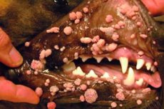
Oral papillomatosis is usually seen in young dogs as stalked, raised masses with a classic fimbriated shape, that is, they are round in shape but often have a rough surface resembling a cauliflower. Such papillomas are defined as exophytic; their transverse size is about 1.5-2 cm. [4]
The first signs of the disease are manifested by one or more small bumps of pink, whitish, grayish or fleshy color on the lips and/or on the mucous membranes of the mouth. Over time, the papillomas enlarge and spread to the mucous membranes of the cheeks, tongue and nose, and in advanced forms can even be found in the pharynx.
Endophytic (inverted) viral warts occur on the body in dogs, most commonly on the belly and paws, and appear as raised plaques of dermal nodules that spread into the skin; the plaque is cup- or dome-shaped with a keratin-filled center. [5]
Papillomatosis of the eyelids in dogs - papillomas of the conjunctiva - can be either in the form of exophytic papillary masses or as squamous cell papillomas of various colors, having a fibrovascular core with slight hyperkeratosis.
Complications and consequences
Traumatizing the papilloma can cause it to become ulcerated and inflamed. [6] In some cases, dogs may develop additional papillomas that gradually increase in size and may spread from the mouth to the entire muzzle.
According to clinical observations, in skin lesions of dogs (especially immunosuppressed animals) with papillomavirus types CPV II-XVII, there is a possibility of malignant transformation of viral papillomas with the development of squamous cell carcinoma. [7]
Diagnostics of the papillomatosis in dogs
The diagnosis of papillomatous lesions is based on the history and clinical picture and is confirmed by histological examination of the sample (biopsy); PCR analysis of blood (or scraping of epithelial cells from the affected area); determining the antigens of papillomavirus IHC (immunohistochemistry), as well as ISH (in situ hybridization) - detection of papillomavirus DNA.
Differential diagnosis
Differential diagnoses include non-CPV-induced squamous cell papillomas (which arise spontaneously from an unknown cause, usually in older dogs); dermal fibroblastic proliferation, infundibular keratoacanthoma, and malignant verruciform epidermodysplasia.
Treatment of the papillomatosis in dogs
Most papillomas in dogs go away on their own, and in mild cases, no treatment is needed.
Nevertheless, to eliminate these skin formations of viral etiology, topical medications can be used: cream with antiviral action Imiquimod (Aldara), ointment Antipapilloma-eco (avoid getting this product on healthy skin), drops Papillox (with celandine extract). For conjunctival papillomas, veterinary Forvet eye drops are used.
To activate immunity, veterinarians recommend the drug Fosprenyl: the solution can be taken internally, as well as administered intramuscularly.
Papillomas formed in the dog's mouth may be subject to secondary infection by bacteria, and then require broad-spectrum antibiotics, most often used macrolide antibiotic Summamed or Azithromycin for papillomatosis (in the form of injections, the course of treatment - 10 days).
When a dog has a large number of papillomas that make eating difficult, resort to removing them using traditional surgery, electrosurgery or cryosurgery.
Prevention
The main way to prevent papillomatosis in dogs is to avoid contact with infected animals and strengthen the immune system (by supplementing the diet with vitamin supplements).
Forecast
The prognosis is usually good, as most oral COPV lesions spontaneously regress without intervention, due to the development of a cell-mediated immune response.
Squamous cell papillomas, on the other hand, do not disappear, but usually do not enlarge.