Medical expert of the article
New publications
Fetal pyeloectasia of the kidneys
Last reviewed: 29.06.2025

All iLive content is medically reviewed or fact checked to ensure as much factual accuracy as possible.
We have strict sourcing guidelines and only link to reputable media sites, academic research institutions and, whenever possible, medically peer reviewed studies. Note that the numbers in parentheses ([1], [2], etc.) are clickable links to these studies.
If you feel that any of our content is inaccurate, out-of-date, or otherwise questionable, please select it and press Ctrl + Enter.

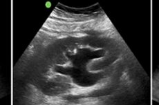
Fetal renal pyeloectasia may be detected when the collecting renal mechanism is evaluated. The problem is an increase in the anteroposterior size of the renal pelvis due to the accumulation of urinary fluid. This pathology is spoken of as an independent (physiological) disorder, or a concomitant process on the background of urological diseases accompanied by urodynamic disorders. Pyeloectasia is detected in the course of ultrasonic diagnosis. Treatment is not always required: the need for therapeutic measures is determined individually. [1]
Epidemiology
Urinary tract anomalies are diagnosed in 5% of newborn infants. They account for 25% of all intrauterine congenital anomalies, and such defects account for about 4% of perinatal infant mortality. The most common disorder, which is detected at the antenatal ultrasound stage, is pyeloectasia, often bilateral or left-sided.
The problem is detected during an ultrasound scan between the 18th and 22nd week of gestation. It occurs in about 2% of cases. Pyeloectasia in a boy fetus is detected on average 4 times more often than in girls, which can be explained by the peculiarities of the anatomy of the male urogenital system. The final determination of the degree of enlargement of the renal pelvis in the fetus is carried out by ultrasound examination at 32 weeks of the gestational period. [2]
Causes of the fetal pyeloectasia of the kidneys
Physiologic pyeloectasia in the fetus is often transient and is due to stenosis of the urinary tract, but often the pathology develops due to congenital abnormalities in the formation of the urinary system. This can be abnormalities in the development of the kidneys, urethra, ureters. Defects arise mainly due to genetic abnormalities, but the problem can also be provoked by the wrong lifestyle of a pregnant woman: a special unfavorable role is played by smoking, drinking alcoholic beverages, etc. Another possible cause is the narrowing of the lumen of the urethra with the formation of so-called strictures. Such a problem can only be eliminated surgically.
Congenital causes of renal pyeloectasia formation come in dynamic and organic.
Dynamic causes include the following:
- Narrowing (stenosis) of the external urethral opening;
- Severe narrowing of the foreskin in boys;
- Urethral strictures;
- Neurogenic disorders of bladder function.
Possible organic causes:
- Kidney developmental defects that cause compression of the ureter;
- Developmental defects in the walls of the upper urinary system;
- Developmental defects in the ureter;
- Defects in the blood network supplying the upper urinary system.
Fetal renal pyeloectasia is formed under the influence of various developmental anomalies and genetic factors. Such risk factors may play a role in the occurrence of the problem:
- Unfavorable ecology, increased radiation background;
- Narrowing of the urinary ducts;
- Hereditary predisposition, inflammatory diseases, pre-eclampsia, pyeloectasia in the future mother;
- Developmental defects in any part of the genitourinary system;
- An incomplete urethral valve;
- Ureteral blockage.
Fetal pyeloectasia on both sides, bilateral pathology is relatively rare and in many cases disappears after the baby's first urination.
Intrauterine disorder is provoked by the following factors:
- Urethrocele is an abnormal urine outflow due to a blockage (stenosis) of the ureter's entrance to the bladder;
- Ectopia - defective insertion of the ureter not into the bladder, but into the vaginal vestibule (thus forming pyeloectasia in a girl fetus), prostate gland, seminal canal or seminal vesicles (in boys);
- Megaloureter is an abnormally dilated ureter that prevents it from emptying normally;
- Hydronephrosis - progressive enlargement of the renal pelvis and cups, leading to impaired urinary outflow.
Pathogenesis
The term "pyeloectasis" is derived from the Greek words "pyelos", "pelvis", and "ectasia", "enlargement". Sometimes not only the pelvis, but also the calyxes are dilated: in such a case we are talking about pyelocalicectasia or hydronephrotic change. If the pelvis and ureter are dilated, then we talk about ureteropyeloectasia, or megoureter.
The pelvis dilates due to increased intrarenal urine pressure due to an obstruction in the urine flow pathway. The problem may be due to either backflow from the bladder, narrowing of the urinary tract below the pelvis, or increased urethral pressure.
In many children, the ureter is narrowed in the area where the pelvis enters the ureter, or where the ureter enters the bladder. It can also be due to underdevelopment of the organ, or compression of the ureter by adhesions, neoplasm, vessel, etc. A formed valve in the area of the pelvic-ureteric junction is somewhat less often the "culprit".
The most common underlying cause of pyeloectasia is considered to be uretero-ureteral reflux. The essence is that normally the development of such reflux is prevented by the valve system, which is present in the area of the ureter's entrance to the bladder. In the case of reflux, this system does not function, so the urine in the process of bladder contraction is directed upward instead of downward.
It is important to realize that pyeloectasia is not an independent pathology, but only an indirect manifestation of impaired urine flow from the pelvis due to some defect in structure, infectious process, reflux movement of urine, etc.
During the intrauterine period and during periods of intense growth, it is important to monitor changes in the size of the renal pelvis. The frequency of such monitoring depends on each specific case and is determined individually by the specialist.
Since the kidneys are paired organs, pyeloectasia can be unilateral or bilateral (affecting one or both kidneys). Pathology can be the result of an infectious process in the urinary tract, or it can provoke the development of inflammatory disease itself.
In newborn infants with general immaturity (e.g. Prematurity), pelvic dilatation often disappears on its own as the organs and systems mature. In such situations, the term pyeloectasia is often replaced by "pelvic atony" or "hypotonia".
Any cases of pelvic dilatation require regular and mandatory monitoring by a nephrologist and ultrasound technician, even though in most children the problem is transient and disappears with time. [3]
Symptoms of the fetal pyeloectasia of the kidneys
In many cases, renal pyeloectasia has an asymptomatic course. But this does not always happen. About every second child has pain: intense, localized in the lumbar region - in the projection of the kidney in which there is a violation. With bilateral dilatation of the renal pelvis, the pain has a shingling character.
Sometimes pyeloectasia can cause an attack of renal colic. This is a very painful condition that is accompanied by severe pain syndrome, up to the development of painful shock. Especially unfavorable course of such an attack is noted in infants, it requires urgent differential diagnosis with urolithiasis and pyelonephritis.
Another common symptom is urinary dysfunction, more commonly pollakiuria (ineffective urges to urinate). Urinary fluid does not come out in a stream, but drips or does not come out at all. Additional but relatively rare symptoms of pyeloectasia include pain during urination, tearing and burning.
Newborn babies may show such early signs as fever and general lethargy, which is a consequence of general intoxication of the body. Often such symptomatology indicates a bilateral disorder.
Since the kidneys are involved in the regulation of blood pressure in the arteries, a significant increase in blood pressure is possible in pyeloectasia. The cause of this phenomenon is a disorder of the glomerular filtration.
In later stages, signs of insufficient renal function may develop.
In general, the symptomatology in most cases is either absent or presented by one or two symptoms. Therefore, an objective comprehensive examination is always used to correctly diagnose renal pyeloectasia. [4]
Complications and consequences
Almost any pathology is capable of causing the development of complications, and renal pyeloectasia is no exception. Complications may be associated with deterioration of organ function, development of inflammatory reactions or tissue atrophic processes, formation of renal sclerosis:
- Renal failure - a sharp or gradually increasing disorder of filtration, and secretory-excretory capacity of the kidneys. Pathology proceeds with a violation of water-electrolyte equilibrium, increasing intoxication and subsequent dysfunction of internal organs.
- Pyelonephritis is a nonspecific infection and inflammation characterized by simultaneous or sequential involvement of the pelvic and renal parenchyma.
- Atrophic processes in the renal parenchyma are a reduction in the size of the affected tissue with impairment or cessation of organ function.
- Nephrosclerosis is a secondary renal pathology, the essence of which lies in the reduction in the size of the organ and the gradual replacement of structures with fibrous tissue. The process is caused by the gradual death of nephrons.
Diagnostics of the fetal pyeloectasia of the kidneys
Fetal renal pyeloectasia is diagnosed by an obstetrician-gynecologist during routine screening during pregnancy. After the baby is born, repeated diagnostic measures are performed to determine the cause of pelvic dilatation and the presence of functional disorders of the kidneys. In many cases, a wait-and-see approach is used, ultrasound is repeated approximately every two months. If the results of this observation show a worsening of the condition, then additional diagnostics is prescribed to specify the etiology of the disease. [5]
To assess kidney functionality in a newborn baby, these laboratory tests are prescribed:
- Urinalysis;
- Zimnitzky's analysis;
- Nechiporenko's analysis;
- Rehberg test;
- Addis-Kakowski test;
- Blood ionogram;
- Blood chemistry (creatinine, urea).
These tests will help the doctor understand how much kidney function is impaired or prove that the organs continue to work normally despite pyeloectasia.
If the laboratory diagnosis reveals an inflammatory process, then additionally prescribe a bacteriological culture of urine to identify the causative agent.
Instrumental diagnosis in this disorder is considered the most informative and helps to clearly identify the cause of pathologically enlarged pelvis and renal cups. With the help of instrumental methods, specialists determine the size, location, shape of the kidneys and the severity of pyeloectasia. When performing an ultrasound examination, the size of the left and right pelvis is determined at least twice.
Mandatory ultrasound diagnosis is carried out in the prescribed order in the second half of the gestational period, from 17 to 22 weeks, in order to identify possible abnormalities in the development of the future baby. Fetal pyeloectasia on ultrasound is determined if the size of the renal pelvis exceeds the norm:
- In the second trimester, 4 to 5 millimeters;
- In the third trimester, 7 millimeters.
A small deviation within 1 mm is a moderate pyeloectasia, which is very likely to disappear in the future. But the size of the enlargement of more than 10 mm is a serious disorder that requires immediate medical consultation. [6]
Differential diagnosis
In the case of moderate pyeloectasia, the newborn infant undergoes regular ultrasound examinations every 2-3 months. If a urinary infection joins, or pelvic dilatation progresses, then a complete urologic diagnosis is performed, including radiologic procedures such as cystography, excretory urography, radioisotope renal study. Such measures help to carry out differential diagnosis and determine the correct diagnosis - to find out the degree and cause of urinary flow disorder, justify and prescribe therapy.
Pyeloectasia is an incomplete diagnosis. It is important to recognize the cause of renal pelvic dilatation, for which a differential diagnosis is mandatory. Here are some examples of pathologies that require special attention of the doctor in terms of confirmation or exclusion:
- Hydronephrosis due to an obstruction at the transition from the pelvis to the ureter.
- Uretero-ureteral reflux, which is a backflow of urinary fluid from the bladder to the kidney.
- Megaureter, characterized by a sharply dilated ureter.
- Posterior urethral valves in boys.
- Ureteral ectopia, in which the ureter does not flow into the bladder but into the vagina in girls or into the urethra in boys.
- Ureterocele, accompanied by bloating of the ureter in the area where it enters the bladder, and narrowing in the area of the exit orifice.
Additional differentiation is made with pyelonephritis and urolithiasis. [7]
Who to contact?
Treatment of the fetal pyeloectasia of the kidneys
Doctors do not have a single and universal scheme of treatment of pyeloectasia: the tactics depend on the characteristics of the enlargement, and the dynamics of the process, as well as on the diagnosed or suspected cause of the disorder.
For example, in cases of severe kidney dysfunction and impairment, surgical intervention may be required to remove the obstruction to urine flow. In such situations, a wait-and-see approach can cause irreparable damage to the child's body.
If there is no strong expansion and visible impairment of kidney function, there are no negative dynamics (according to the results of ultrasound and laboratory tests), then it is optimal to apply observation and conservative therapy. It consists of physiotherapeutic procedures, taking herbal remedies, control ultrasound.
If pyeloectasia in a baby runs without symptoms, then among the main recommendations will be regular ultrasound monitoring, proper nutrition (without stress on the kidneys) and prevention of inflammatory processes in the urogenital apparatus. If the problem begins to progress, then additionally prescribed drugs that facilitate the outflow of urine and stop the inflammatory reaction.
Such medications may be used to eliminate the infection and inflammatory response:
- Broad-spectrum antibiotics with antibacterial activity;
- Uroseptic;
- Immunomodulators;
- Multivitamin preparations;
- Litholytics (medicines that prevent crystals from forming and settling).
Surgical intervention can correct the defect and eliminate urethral reflux. The surgery is often performed using endoscopic methods, avoiding open intervention, using mini-instruments that are inserted through the urethra. [8]
The following surgical practices may be utilized:
- Plastic surgery of the uretero-pelvic segment and excision of the distended pelvic sheath with ureteral reinsertion into the kidney, bouching, balloon dilation and endotomy with laser or electrocurrent treatment;
- Palliative intervention and normalization of urinary outflow in acute inflammatory process with epicystostomy, nephrostomy, stent catheter placement;
- Removal of a tumor process that is impairing urodynamics;
- Nephrectomy for renal dysfunction and destruction of kidney tissue (children undergo surgery only if more than 90% of the kidney tissue is affected).
In case of moderate dilatation of the pelvis may be recommended to take a decoction of diuretic herbs and herbal uroseptic.
Prevention
Preventive measures to prevent the development of fetal renal pyeloectasia include:
- Timely referral to doctors and treatment of various disorders of the genitourinary apparatus;
- Normalizing the drinking regimen;
- Compliance with all recommendations for a healthy lifestyle, avoiding bad habits during pregnancy.
The birth of a child is a wonderful event, but it is important to prepare for it even before conception. To prevent the development of congenital abnormalities, including fetal renal pyeloectasia, women should follow these recommendations:
- Sanitize chronic infectious foci - including tonsillitis, pyelonephritis, sinusitis, etc. During gestation, such diseases are often aggravated, which can adversely affect the formation of the body of the future baby.
- Additionally examine for certain infectious pathologies, such as herpesvirus and cytomegalovirus infection, chlamydia, ureaplasmosis, toxoplasmosis. Many pathogens become factors in the development of birth defects in the fetus. If such an infection is detected, it is necessary to undergo an appropriate course of treatment.
- Eradicate all bad habits, avoid alcoholic beverages throughout the entire process of carrying.
- Correct hormonal abnormalities, if any were detected in preparation for pregnancy.
- Eat a full and balanced diet, paying special attention to foods rich in folic acid (tomatoes, liver, beans, spinach). If necessary, the doctor may prescribe additional multivitamin preparations.
Preparing for pregnancy should be done at least a few months before the intended conception.
Forecast
In most children, moderate pyeloectasia disappears on its own due to the maturation of the urinary apparatus and, in particular, the kidneys after birth. Only in a few cases there is still a need for therapeutic measures.
In general, the prognosis depends on the severity and underlying cause of the disorder. Children with moderate to moderate pyeloectasia should be regularly monitored and treated as necessary. In such situations, there is every chance to wait for the elimination or significant reduction in the severity of the defect.
Today, no specialist can confidently predict the behavior and outcome of fetal pyeloectasia of the kidneys. The question of the necessity and completeness of treatment is decided only after the arrival of the baby into the world, as well as in the process of further observation and diagnosis. In case of severe enlargement of the pelvis and gradual aggravation of the pathology, associated with deterioration of the functional state of the organs, surgical intervention may be recommended.
List of authoritative books and studies related to the study of fetal renal pyeloectasia
- "Pediatric Urology: Surgical Complications and Management" - by Ciro Esposito, Alaa El-Ghoneimi (Year: 2008)
- "Pediatric Urology: Evidence for Optimal Patient Management" - by John G. Gearhart (Year: 2013)
- "Clinical Pediatric Urology" - by John P. Gearhart, Richard C. Rink (Year: 2006)
- "Pediatric Urology" - by Douglas Canning, M. Chad Wallis (Year: 2010)
- "Clinical Urography" - by Arthur C. Baert (Year: 2013)
- "Urology in Childhood" - by Arthur L. Burnett, John P. Gearhart (Year: 2008)
- "Urology: Pediatric Urology" by John G. Gearhart, Richard C. Rink (Year: 2001)
- "Atlas of Pediatric Urologic Surgery" - by Frank H. Netter, Lane S. Palmer (Year: 2011)
- "Pediatric Robotic Urology" - by Mohan S. Gundeti, Prasad P. Godbole (Year: 2017)
- "Pediatric Urology: Current Clinical Urology" - by Stephen A. Zderic (Year: 2010)
Literature
Volodin, N. N. Neonatology / Antonov A. G.. Arestova N. N. Baibarina E. N. N., Baibarina E. Et al. / Edited by N. N. Volodin - Moscow: GEOTAR-Media, 2009.

