Medical expert of the article

New publications
Ectopic pregnancy
Last reviewed: 04.07.2025

All iLive content is medically reviewed or fact checked to ensure as much factual accuracy as possible.
We have strict sourcing guidelines and only link to reputable media sites, academic research institutions and, whenever possible, medically peer reviewed studies. Note that the numbers in parentheses ([1], [2], etc.) are clickable links to these studies.
If you feel that any of our content is inaccurate, out-of-date, or otherwise questionable, please select it and press Ctrl + Enter.
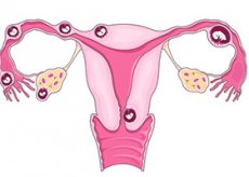
In a normal pregnancy, the fertilized egg travels down the fallopian tube toward the uterus, where it attaches to the wall and begins to grow. But in a condition called an ectopic pregnancy, the fertilized egg does not make it to the uterus, but begins to grow elsewhere, often in the fallopian tube. This is why it is often called a tubal ectopic pregnancy.
In rare cases, the egg attaches to the ovary, abdominal muscles, or cervical canal. It is impossible to save the fetus in such a pregnancy. If the egg begins to grow in the fallopian tube, the tube may be damaged or ruptured, which can lead to severe bleeding, which can even be fatal. If you have been diagnosed with an ectopic pregnancy, it must be terminated immediately before complications develop.
Epidemiology
The incidence of ectopic pregnancy in the United States has more than quadrupled and is now 20 per 1,000 pregnancies.
Ectopic pregnancy accounts for 10% of pregnancy-related deaths in women in the United States. Most deaths are due to bleeding and are potentially preventable.
Over the last decade, there has been a clear trend towards an increase in the frequency of ectopic pregnancy. This fact can be explained in two ways. On the one hand, the prevalence of inflammatory processes in the internal genital organs is constantly growing; the number of surgical interventions on the fallopian tubes, which are carried out to regulate childbearing, is increasing; the number of women using intrauterine and hormonal methods of contraception is growing; ovulation inducers are increasingly being introduced into the practice of treating infertility. On the other hand, diagnostic capabilities have improved in recent years, allowing the detection of intact and even regressing ectopic pregnancy.
Currently, ectopic pregnancy occurs in 0.8 to 2.4 cases per 100 women who have given birth. In 4-10% of cases, it is repeated.
Causes ectopic pregnancy
An ectopic pregnancy often occurs as a result of damage to the fallopian tubes. The fertilized egg cannot reach the uterus and is therefore forced to attach to the wall of the tube.
Provocateurs of ectopic pregnancy:
- Smoking (the more you smoke, the higher the risk of ectopic pregnancy).
- Pelvic inflammatory disease (resulting from chlamydia or gonorrhea) that causes scar tissue to form in the fallopian tubes.
- Endometriosis, which causes scar tissue to form in the fallopian tubes.
- Exposure to synthetic estrogen (diethylstilbestrol) before birth.
- Previous ectopic pregnancy in the fallopian tubes.
Certain medical interventions may increase the risk of ectopic pregnancy:
- Surgeries on the fallopian tubes in the pelvic area (tubal ligation) or to remove scar tissue.
- Treatment of infertility.
Ectopic pregnancy is associated with taking medications to ovulate more eggs. Scientists do not yet know whether ectopic pregnancy is caused by hormones or damage to the fallopian tubes.
If you are pregnant and are concerned about an ectopic pregnancy, you should be thoroughly examined. Doctors do not always agree on the risk factors for ectopic pregnancy, but one thing is clear - the risk increases after a history of ectopic pregnancy, surgery on the fallopian tubes, or pregnancy with an IUD.
Pathogenesis
Implantation of the fertilized egg outside the uterine cavity may occur due to a disruption of the transport function of the fallopian tubes, as well as due to a change in the properties of the fertilized egg itself. Combinations of both causal factors in the development of an ectopic pregnancy are possible.
Fertilization of the egg by the sperm under normal conditions occurs in the fimbrial section of the ampulla of the fallopian tube. Due to the peristaltic, pendulum-like and turbulent movements of the tube, as well as due to the flickering of the ciliated epithelium of the endosalpinx, the fragmenting fertilized egg reaches the uterine cavity in 3-4 days, where the blastocyst can remain in a free state for 2-4 days. Then, having lost the shiny shell, the blastocyst plunges into the endometrium. Thus, implantation occurs on the 20th-21st day of the 4-week menstrual cycle. Disruption of the transport function of the fallopian tubes or accelerated development of the blastocyst can lead to implantation of the fertilized egg proximal to the uterine cavity.
Practice shows that dysfunction of the tube is most often associated with inflammatory processes of any etiology. The predominant role is played by non-specific infection, the spread of which is facilitated by abortions, intrauterine contraception, intrauterine diagnostic interventions, complicated course of labor and the postpartum period, and appendicitis. In recent years, a high frequency of chlamydial infection has been revealed in women operated on for ectopic pregnancy. Along with the inflammatory nature of the disorder of the structure and function of the fallopian tubes, the role of endometriosis is extremely important.
The importance of surgical interventions on the fallopian tubes in the structure of causal factors leading to the occurrence of ectopic pregnancy is constantly increasing. Even the introduction of microsurgery does not eliminate such a danger.
Contractile activity of the tube is closely related to the nature of the hormonal status of the body. An unfavorable hormonal background in women can be caused by a violation of the regulation of the menstrual cycle of any nature, age, as well as the use of exogenous hormonal drugs that contribute to the violation or induction of ovulation.
Inadequacy of blastocyst development to the physiological implantation site is associated with excessive biological activity of the egg itself, leading to accelerated formation of trophoblast and possible nidation, not reaching the uterine cavity. It is almost impossible to determine the reason for such rapid blastocyst development.
In some cases, the disruption of the transport of the fertilized egg can be explained by the peculiarities of its path, for example, external migration of the egg after surgical intervention on the appendages: the egg from the only ovary through the abdominal cavity gets into the only tube on the opposite side. Cases of transperitoneal migration of spermatozoa in some malformations of the internal genital organs have been described.
In recent years, there have been reports of the possibility of tubal pregnancy after in vitro fertilization and blastocyst transfer into the uterus.
The tube, ovary, abdominal cavity, and even the rudimentary horn of the uterus do not have a powerful, specifically developed mucous and submucous membrane, characteristic of physiological pregnancy. Progressive ectopic pregnancy stretches the fetal receptacle, and the chorionic villi destroy the underlying tissue, including blood vessels. Depending on the location of the pregnancy, this process can proceed faster or slower, accompanied by more or less bleeding.
If the fertilized egg develops in the isthmic section of the tube, where the height of the mucous membrane folds is small, the so-called basotropic (main) growth of the chorionic villi takes place, which quickly destroy the mucous, muscular and serous layers of the tube, and after 4-6 weeks this leads to perforation of the wall with destruction of the vessels, powerfully developed in connection with pregnancy. Termination of pregnancy occurs according to the type of external rupture of the fetal receptacle, i.e. rupture of the pregnant tube, which is accompanied by massive bleeding into the abdominal cavity. The same mechanism is used for termination of pregnancy localized in the interstitial section of the tube. However, due to the significant muscular layer surrounding this section of the tube, the duration of pregnancy can be longer (up to 10-12 weeks or more). Blood loss due to the extremely developed blood supply to this area during rupture of the fetal receptacle is usually massive.
The integrity of the mesenteric edge of the tube is extremely rarely violated. In this case, the fertilized egg and the gushing blood end up between the sheets of the broad ligament. Casuistic cases have been described where the fertilized egg did not die, but continued to develop interligamentously until a significant time.
In case of ampullar localization of tubal pregnancy, implantation of the fertilized egg into the fold of endosalpinx (columnar or acrotropic attachment) is possible. In this case, the growth of the chorionic villi may be directed towards the lumen of the tube, which 4-8 weeks after nidation is accompanied by a violation of the internal capsule of the fetal receptacle, and this, in turn, leads to slight or moderate bleeding. Antiperistaltic movements of the tubes can gradually expel the detached fertilized egg into the abdominal cavity: a tubal abortion occurs. When the fimbrial section of the tube closes, blood pouring into the lumen of the tube leads to the formation of hematosalpinx. When the lumen of the ampulla is open, blood flowing out of the tube and coagulating in the area of its funnel can form a peritubular hematoma. Repeated, more profuse bleeding leads to the accumulation of blood in the recto-uterine pouch and the formation of a so-called retro-uterine hematoma, separated from the abdominal cavity by a fibrous capsule fused with intestinal loops and the omentum.
In extremely rare cases, the fertilized egg expelled from the tube does not die, but attaches to the parietal or visceral peritoneum of the abdominal organs (most often to the peritoneum of the recto-uterine pouch). A secondary abdominal pregnancy develops, which can exist for different periods of time, up to full-term pregnancy. Even more rarely, the fertilized egg can implant in the abdominal cavity primarily.
Ovarian pregnancy rarely lasts for a long time. Usually there is an external rupture of the fetal receptacle, accompanied by significant bleeding. If the pregnancy develops on the surface of the ovary, such an outcome occurs early. In the case of intrafollicular localization, the interruption occurs later.
Cervical pregnancy is a rare but potentially severe form of ectopic pregnancy due to the high risk of bleeding. Cervical pregnancy is usually treated with methotrexate.
Symptoms ectopic pregnancy
During the first few weeks, an ectopic pregnancy causes the same symptoms as a normal pregnancy: no menstrual periods, fatigue, nausea, and breast tenderness.
The main signs of an ectopic pregnancy:
- Pain in the pelvic or abdominal area, which may be acute and one-sided, but over time may spread to the entire abdominal cavity. The pain increases with movement or strain.
- Vaginal bleeding.
If you think you are pregnant and experience any of the above symptoms, seek medical attention immediately.
The first weeks of an ectopic pregnancy are no different from a normal pregnancy. During this period, the following is observed:
- Absence of menstrual cycle.
- Chest pain.
- Fatigue.
- Nausea.
- Frequent urination.
But if the ectopic pregnancy continues, other symptoms arise, including:
- Pain in the pelvic organs or abdominal cavity (usually by 6-8 weeks after the end of the menstrual cycle). The pain intensifies with movement or strain, can be acute, one-sided, and eventually spreads to the entire abdominal cavity.
- Moderate to heavy vaginal bleeding.
- Pain during sexual intercourse or physical examination by a doctor.
- Pain in the shoulder area due to bleeding into the abdominal area due to irritation of the diaphragm.
The symptoms of an early ectopic pregnancy and a miscarriage are often the same.
Typically, at the beginning of pregnancy, the fertilized egg travels down the fallopian tube toward the uterus, where it attaches to the wall and begins to develop. But in 2% of diagnosed pregnancies, the fertilized egg becomes lodged outside the uterus, resulting in an ectopic pregnancy.
In an ectopic pregnancy, the fetus cannot develop for a long time, but reaches such a size that it leads to a rupture of the tube and bleeding, which can be fatal for the mother. A woman who has symptoms of an ectopic pregnancy requires immediate medical attention. In most cases of ectopic pregnancy, the fertilized egg attaches to the fallopian tube. In rare cases:
- The egg attaches and begins to grow in the ovary, in the cervical canal, or in the abdominal cavity (excluding the reproductive organs).
- One or more eggs develop in the uterus, while another egg (or several) grow in the fallopian tube, cervical canal, or abdominal cavity.
- In very rare cases, the egg begins to develop in the abdominal cavity after the uterus has been removed (hysterectomy).
When to seek medical help?
If you are expecting a baby, watch closely for symptoms that may indicate an ectopic pregnancy, especially if you are predisposed to it.
For vaginal bleeding and acute abdominal pain (before or after diagnosis of pregnancy or during treatment of an ectopic pregnancy):
- call an ambulance;
- go to bed and rest;
- Do not make any sudden movements until your doctor has assessed your health condition.
If you experience persistent minor abdominal pain, contact your doctor.
 [ 19 ]
Observation
To observe means to wait a little and see if the condition improves. But in case of an ectopic pregnancy, due to the risk of death, you cannot stay at home and wait for a miracle. Call an ambulance immediately at the first signs of an ectopic pregnancy.
Specialists to contact
- gynecologist
- family doctor
- emergency doctor
If an ectopic pregnancy is diagnosed, treatment is carried out by a gynecologist.
Forms
By localization |
With the flow |
| tubal (ampullary, isthmic, interstitial); ovarian; abdominal; cervico-isthmic | progressive; tubal abortion; ruptured fallopian tube; frozen |
Unlike ICD-10, in domestic literature tubal pregnancy is divided into:
- ampullary;
- isthmic;
- interstitial.
Interstitial tubal pregnancy accounts for slightly less than 1% of ectopic pregnancies. Patients with interstitial tubal pregnancy in most cases seek medical attention later than with ampullary or isthmic pregnancy. The incidence of pregnancy in the uterine angle increases to 27% in patients with a history of salpingectomy and IVF and PE. Interstitial tubal pregnancy is associated with the majority of deaths due to ectopic pregnancy in general, since it is often complicated by uterine rupture.
Ovarian pregnancy is divided into:
- developing on the surface of the ovary;
- developing intrafollicularly.
Abdominal pregnancy is divided into:
- primary (implantation in the abdominal cavity occurs initially);
- secondary.
Depending on the location of implantation of the fertilized egg, ectopic pregnancy is divided into tubal, ovarian, located in the rudimentary horn of the uterus, and abdominal. Among all cases of tubal pregnancy, depending on the location of the fetal receptacle, there are ampullar, isthmic and interstitial. Ovarian pregnancy can be observed in two variants: developing on the surface of the ovary and inside the follicle. Abdominal ectopic pregnancy is divided into primary (implantation initially occurs on the parietal peritoneum, omentum or any organs of the abdominal cavity) and secondary (attachment of the fertilized egg in the abdominal cavity after its expulsion from the fallopian tube). Ectopic pregnancy in the rudimentary horn of the uterus, strictly speaking, should be attributed to the ectopic variety of uterine pregnancy, but the peculiarities of its clinical course force us to consider this localization in the group of proximal variants of ectopic pregnancy.
Among all types of ectopic pregnancy, it is customary to distinguish between common and rare forms. The former include ampullar and isthmic localization of tubal pregnancy, which account for 93-98.5% of cases. Ampullar localization of tubal pregnancy is somewhat more common than isthmic.
Rare forms of ectopic pregnancy include interstitial (0.4-2.1%), ovarian (0.4-1.3%), and abdominal (0.1-0.9%). Even rarer is ectopic pregnancy developing in the rudimentary horn of the uterus (0.1-0.9%) or in an accessory fallopian tube. Casuistry includes extremely rare cases of multiple pregnancies with various localizations: a combination of uterine and tubal, bilateral tubal, and other combinations of ectopic localization of the ovum.
The localization of the ectopic fetal receptacle is closely related to the features of the clinical course of the disease, among which progressive and impaired forms are distinguished. The pregnancy interruption can occur by the type of external rupture of the fetal receptacle: rupture of the ovary, rudimentary horn of the uterus, interstitial section of the fallopian tube, often - the isthmic section, rarely - the ampullar section. The second type of pregnancy interruption is an internal rupture of the fetal receptacle, or tubal abortion. This type most often occurs in pregnancy interruption located in the ampullar section of the tube. In recent years, due to the improvement of diagnostic capabilities, there has been a tendency to isolate a regressing form of ectopic pregnancy.
[ 20 ], [ 21 ], [ 22 ], [ 23 ]
Abdominal pregnancy
It is considered a rare form of ectopic pregnancy (0.3–0.4%). The localization of abdominal pregnancy varies: omentum, liver, sacrouterine ligaments, rectouterine pouch. It can be primary (implantation occurs in the abdominal organs) and secondary (initially, implantation occurs in the tube, and then, as a result of a tubal abortion, the fertilized egg is expelled from the tube and is reimplanted in the abdominal cavity). This difference is of purely theoretical interest, and the initial implantation can only be established by histological examination, since by the time of surgery the tube is already macroscopically unchanged.
Abdominal pregnancy, both primary and secondary, is extremely rare. Progressive primary pregnancy is practically not diagnosed; its termination gives a picture of a disrupted tubal pregnancy.
Secondary abdominal pregnancy occurs after a tubal abortion or a ruptured tube, and very rarely after a ruptured uterus. An abdominal pregnancy can be carried to a later stage, which poses a serious threat to the woman's life, and the fetus is rarely viable. More than half of the fetuses have developmental defects.
Secondary abdominal pregnancy can be suspected in women who had episodes of lower abdominal pain in the early stages, accompanied by small bloody discharge from the vagina. Typical complaints of women about painful fetal movements. During external examination of the patient, it is possible to detect an abnormal position of the fetus. Its small parts can be clearly palpated. There are no contractions of the fetal receptacle, which are usually determined by palpation. During internal examination, attention should be paid to the displacement of the cervix upward and to the side. In some cases, it is possible to palpate the uterus separately from the fetal receptacle. Ultrasound scanning reveals the absence of the uterine wall around the amniotic sac.
[ 24 ], [ 25 ], [ 26 ], [ 27 ], [ 28 ], [ 29 ]
Ovarian pregnancy
One of the rare forms of ectopic pregnancy, its frequency is 0.1-0.7%. There are two forms of this pregnancy: intrafollicular and epiphoral. In the intrafollicular form, fertilization and implantation occur in the follicle, in the epiphoral form - on the surface of the ovary.
Cervical pregnancy
The incidence ranges from 1 in 2,400 to 1 in 50,000 pregnancies. The risk is thought to be increased by a previous abortion or cesarean section, Asherman syndrome, maternal use of diethylstilbestrol during pregnancy, uterine fibroids, in vitro fertilization, and embryo transfer. Ultrasound signs of cervical pregnancy:
- absence of a fertilized egg in the uterus or a false fertilized egg;
- hyperechogenicity of the endometrium (decidual tissue);
- myometrial heterogeneity;
- hourglass shaped uterus;
- dilation of the cervical canal;
- fertilized egg in the cervical canal;
- placental tissue in the cervical canal;
- closed internal os.
After confirming the diagnosis, the blood type and Rh factor are determined, a venous catheter is inserted, and the patient's written consent for extirpation of the uterus, if necessary, is obtained. All this is caused by the high risk of massive bleeding. There are reports of the effectiveness of intra-amniotic and systemic methotrexate in cervical pregnancy. The diagnosis of cervical pregnancy is often made only during diagnostic curettage for a suspected abortion in progress or incomplete abortion, when profuse bleeding has begun. To stop bleeding, depending on its intensity, tight vaginal tamponade, suturing of the lateral vaginal fornices, application of a circular suture to the cervix, insertion of a Foley catheter into the cervical canal and inflation of the cuff are used. Embolization of bleeding vessels, ligation of the uterine or internal iliac arteries are also used. If all of the above measures are ineffective, extirpation of the uterus is performed.
Pregnancy in the rudimentary horn of the uterus
Occurs in 0.1–0.9% of cases. Anatomically, this pregnancy can be classified as uterine, but due to the fact that in most cases the rudimentary horn has no connection with the vagina, clinically such a pregnancy proceeds as ectopic.
Pregnancy in the rudimentary horn, which has an insufficiently developed muscular layer and an inadequate mucous membrane, occurs under the following conditions: the cavity of the horn communicates with the fallopian tube, the desquamation phase does not occur in the mucous membrane and, therefore, hematometra does not form, which prevents implantation of the fertilized egg. The mechanism of penetration of the blastocyst into the cavity of the rudimentary horn is apparently associated with transperitoneal migration of spermatozoa or egg.
Progressive pregnancy is diagnosed extremely rarely. It can be suspected based on unusual data from an internal gynecological examination: an enlarged uterus (at terms over 8 weeks, inconsistent with the period of delayed menstruation) is deviated to the side; on the opposite side, a tumor-like painless formation of a soft consistency is determined, connected to the uterus by a thick stalk. Ultrasound examination or laparoscopy provide invaluable assistance.
The pregnancy disorder occurs as an external rupture of the fetal receptacle, accompanied by profuse bleeding and requiring emergency surgical intervention. The scope of the operation in typical cases is the removal of the rudimentary horn together with the adjacent fallopian tube.
Intraligamentary pregnancy
It accounts for 1 in 300 cases of ectopic pregnancy. It usually occurs secondarily, when the fallopian tube ruptures along the mesenteric edge and the ovum penetrates between the layers of the broad ligament. Intraligamentary pregnancy is also possible with a fistula connecting the uterine cavity and parametrium. The placenta can be located on the uterus, bladder, or pelvic wall. If it is impossible to remove the placenta, it is left. There are reports of successful delivery of full-term intraligamentary pregnancies.
Rare variants of ectopic pregnancy
Combination of intrauterine and ectopic pregnancy
The frequency, according to different authors, is from 1 in 100 to 1 in 30,000 pregnancies. It is higher after ovulation induction. Having identified the fertilized egg in the uterus, the second fertilized egg is often ignored during ultrasound. The results of multiple studies of the beta subunit of hCG do not differ from those in normal pregnancy. In most cases, an operation is performed for an ectopic pregnancy and the uterine pregnancy is not interrupted. It is also possible to introduce potassium chloride into the fertilized egg located in the fallopian tube (during laparoscopy or through the lateral vaginal fornix). Methotrexate is not used.
[ 30 ], [ 31 ], [ 32 ], [ 33 ]
Multiple ectopic pregnancy
It is even less common than a combination of intrauterine and extrauterine pregnancy. There are many known variants of the number and location of the fertilized eggs. About 250 cases of ectopic twin pregnancy have been described. In most cases, these are ampullary or isthmic tubal pregnancies, but ovarian, interstitial tubal, and abdominal pregnancies have also been described. Ectopic twin and triplet pregnancies are possible after resection of the fallopian tube and EP. Treatment is the same as for singleton pregnancies.
Pregnancy after hysterectomy
The rarest type of ectopic pregnancy is pregnancy after vaginal or abdominal hysterectomy. Embryo implantation in the fallopian tube occurs shortly before or on the first day after surgery. Ectopic pregnancy is possible at any time after surgery if there is communication between the abdominal cavity and the stump of the cervix or vagina.
Chronic ectopic pregnancy
This is a condition when the fertilized egg does not fully organize after death, and viable chorionic villi remain in the fallopian tube. Chronic ectopic pregnancy occurs when treatment has not been carried out for some reason. Chorionic villi cause repeated hemorrhages in the wall of the fallopian tube, it gradually stretches, but usually does not rupture. With chronic ectopic pregnancy, 86% of patients report pain in the lower abdomen, 68% - bloody discharge from the genital tract. Both symptoms are observed in 58% of women. In 90% of patients, menstruation is absent for 5-16 weeks (on average 9.6 weeks), almost all have a volumetric formation in the small pelvis. Occasionally, with chronic ectopic pregnancy, compression of the ureters or intestinal obstruction occurs. The most informative method for diagnosing chronic ectopic pregnancy is ultrasound. The serum concentration of the β-subunit of hCG is low or normal. Salpingectomy is indicated. The accompanying aseptic inflammation leads to an adhesion process, and therefore the ovary often has to be removed along with the fallopian tube.
[ 34 ], [ 35 ], [ 36 ], [ 37 ], [ 38 ], [ 39 ]
Spontaneous recovery
In some cases, the ectopic pregnancy stops developing and the fertilized egg gradually disappears, or a complete tubal abortion occurs. Surgical treatment is not required. The frequency of such an outcome of ectopic pregnancy and the conditions predisposing to it are unknown. It is also impossible to estimate its prognosis. The content of the β-subunit of hCG cannot serve as a guide.
Persistent ectopic pregnancy
Observed after organ-preserving operations on the fallopian tubes (salpingotomy and artificial tubal abortion). Histological examination usually shows no embryo, and chorionic villi are found in the muscular layer. Implantation occurs medially from the scar on the fallopian tube. Implantation of chorionic villi in the abdominal cavity is possible. Recently, the incidence of persistent ectopic pregnancy has increased. This is explained by the widespread use of organ-preserving operations on the fallopian tubes. Characteristically, there is no decrease in the beta subunit of hCG after surgery. It is recommended to determine the beta subunit of hCG or progesterone on the 6th day after surgery and then every 3 days. The risk of persistent ectopic pregnancy depends on the type of surgery, the initial concentration of the beta subunit of hCG, the gestational age and the size of the ovum. A delay in menstruation of less than 3 weeks and a fetal sac diameter of less than 2 cm increase the risk of persistent ectopic pregnancy. In persistent ectopic pregnancy, both surgical (repeated salpingotomy or, more often, salpingectomy) and conservative treatment (methotrexate) are performed. Many authors prefer conservative treatment, since chorionic villi may be located not only in the fallopian tube and, therefore, are not always detected during repeated surgery. In case of hemodynamic disturbances, surgery is indicated.
Complications and consequences
An ectopic pregnancy can rupture the fallopian tube, reducing the chances of a subsequent pregnancy.
An ectopic pregnancy should be diagnosed early for the safety of the woman and to prevent severe bleeding. A perforated ectopic pregnancy requires immediate surgery to stop severe bleeding in the abdominal cavity. The ruptured fallopian tube is removed completely or partially.
[ 40 ]
Diagnostics ectopic pregnancy
If you suspect you are pregnant, buy a pregnancy test or take a urine test. To determine if you have an ectopic pregnancy, your doctor will:
- will conduct an examination of the pelvic organs to determine the size of the uterus and the presence of formations in the abdominal cavity;
- will prescribe a blood test to detect the pregnancy hormone (the test is repeated after 2 days). In the early stages of pregnancy, the level of this hormone doubles every two days. A low level indicates an anomaly - an ectopic pregnancy.
- Ultrasound shows an image of internal organs. The doctor diagnoses pregnancy at 6 weeks from the last menstrual cycle.
In most cases, an ectopic pregnancy can be determined through a vaginal examination, ultrasound, and blood test. If you have symptoms of an ectopic pregnancy, you should:
- undergo a vaginal examination, during which the doctor will determine pain in the uterus or fallopian tubes, and an increase in the size of the uterus greater than usual;
- have an ultrasound (transvaginal or abdominopelvic), which provides a clear image of the organs and their structure in the lower abdomen. Transvaginal examination (ultrasound) is a more reliable way to diagnose pregnancy, which can be determined as early as 6 weeks after the last menstrual period. In the case of an ectopic pregnancy, the doctor will not see signs of an embryo or fetus in the uterus, but a blood test will show elevated hormone levels.
- Have your blood tested two or more times to measure hormone levels (human chorionic gonadotropin) 48 hours apart. During the first weeks of a normal pregnancy, the level of this hormone doubles every two days. Low or slightly rising levels indicate an ectopic pregnancy or miscarriage. If the level of this hormone is too low, additional tests should be done to determine the cause.
Laparoscopy is sometimes used to detect ectopic pregnancies, which can be seen and terminated as early as 5 weeks. However, it is not used often because ultrasound and blood tests are more accurate.
The main complaints of patients with ectopic pregnancy:
- delayed menstruation (73%);
- bloody discharge from the genital tract (71%);
- pain of varying nature and intensity (68%);
- nausea;
- irradiation of pain to the lumbar region, rectum, inner thigh;
- a combination of three of the above symptoms.
Laboratory and instrumental studies in ectopic pregnancy
The most informative methods for diagnosing ectopic pregnancy are: determination of the concentration of the β-subunit of human chorionic gonadotropin (hCG) in the blood, ultrasound and laparoscopy.
For early diagnosis the following is carried out:
- transvaginal ultrasound;
- determination of the content of the β-subunit of hCG in blood serum.
The combination of transvaginal ultrasound and determination of the concentration of the β-subunit of hCG allows diagnosing pregnancy in 98% of patients from the 3rd week of pregnancy. Ultrasound diagnostics of ectopic pregnancy includes measuring the thickness of the endometrium, sonohysterography, color Doppler. Pregnancy in the uterine angle can be suspected with uterine asymmetry, asymmetric position of the ovum, detected by ultrasound.
The main criteria for ultrasound diagnosis of ectopic pregnancy:
- heterogeneous appendage structures and free fluid in the abdominal cavity (26.9%);
- heterogeneous adnexal structures without free fluid (16%);
- ectopically located fertilized egg with a living embryo (there is a heartbeat) (12.9%);
- ectopic location of the embryo (no heartbeat) (6.9%).
Based on the results of ultrasound, there are 3 types of echographic images of the uterine cavity during ectopic pregnancy:
- I - endometrium thickened from 11 to 25 mm without signs of destruction;
- II - the uterine cavity is enlarged, the anteroposterior size is from 10 to 26 mm, the contents are mainly liquid, heterogeneous due to hematometra and gravid endometrium rejected to varying degrees;
- III - the uterine cavity is closed, M-echo in the form of a hyperechoic strip from 1.6 to 3.2 mm (Kulakov V.I., Demidov V.N., 1996).
To clarify the diagnosis of tubal pregnancy, disrupted by the type of internal rupture of the fetal receptacle, there are numerous additional research methods. The most informative and modern are the following:
- Determination of human chorionic gonadotropin or its beta subunit (beta-chorionic gonadotropin) in blood serum or urine.
- Ultrasound scanning.
- Laparoscopy.
Currently, there are many methods for determining human chorionic gonadotropin. Some of them (for example, biological ones) have lost their leading role. Due to high specificity and sensitivity, preference is given to the radioimmunological method of quantitative determination of B-chorionic gonadotropin in blood serum. Enzyme immunoassay methods for detecting human chorionic gonadotropin in urine, as well as other types of immunological tests (capillary, plate), have earned a positive assessment. Such widely known serological methods for determining human chorionic gonadotropin in urine as the reaction of inhibition of erythrocyte agglutination or sedimentation of latex particles have the right to exist. All laboratory methods for diagnosing pregnancy are highly specific: correct answers are observed from 92 to 100% already from the 9th-12th day after fertilization of the egg. However, they only establish the fact of pregnancy without specifying its localization, so they can be used for. conducting a differential diagnosis with an inflammatory process in the appendages, ovarian apoplexy, endometriosis of the appendages and similar diseases.
Ultrasound examination (US) is a widely used non-invasive method, which in combination with the determination of beta-chorionic gonadotropin can provide high diagnostic accuracy. The main signs of tubal abortion, identified by ultrasound, include the absence of a fertilized egg in the uterine cavity, enlarged appendages, and the presence of fluid in the recto-uterine pouch. Pulsation of the embryonic heart during ectopic pregnancy is rarely recorded.
Transvaginal ultrasound allows to detect the fertilized egg in the uterine cavity when the concentration of beta-chorionic gonadotropin in the blood serum is 1000-1200 IU/L (approximately 5 days after the beginning of the last menstruation). Using transabdominal ultrasound, the fertilized egg in the uterine cavity can be detected when the concentration of beta-chorionic gonadotropin in the blood serum is more than 6000 IU/L.
The most informative method, allowing almost 100% accuracy of differential diagnosis, is laparoscopy. The high assessment of the diagnostic capabilities of laparoscopy is somewhat reduced by the fact that this method is aggressive, it cannot be used in all patients, since complications are possible in the process of its implementation.
Contraindications to laparoscopy are cardiac and pulmonary insufficiency; all types of shock, peritonitis; intestinal obstruction; all diseases and conditions accompanied by impaired blood clotting; adhesions in the abdominal cavity; flatulence; obesity; the presence of infectious diseases. Serious complications rarely accompany laparoscopy. The most common are damage to the small and large intestine, omentum, blood vessels, as well as emphysema of the abdominal wall, omentum and mediastinum. Therefore, to this day, the opinion remains relevant that endoscopy should be performed as the final stage of the examination.
A method well known to gynecologists, such as puncture of the utero-rectal cavity of the abdominal cavity, carried out through the posterior vaginal fornix, has not lost its significance. Obtaining liquid dark blood with small clots confirms the presence of tubal pregnancy. However, it should be remembered that the absence of blood in the puncture does not allow making a categorical conclusion.
In many cases, differential diagnosis is aided by histological examination of endometrial scrapings. The absence of chorionic villi in the presence of decidual transformations of the mucous membrane or other more subtle changes in the endometrium (structures of reverse development of the mucous membrane after pregnancy failure, tangles of spiral vessels, transformation of the uterine epithelium in the form of the Arias-Stella phenomenon and Overbeck's "light glands") most often indicates an ectopic pregnancy.
In difficult to diagnose cases, hysterosalpingography with the introduction of water-soluble contrast agents or its variation - selective salpingography after preliminary catheterization of the fallopian tubes during hysteroscopy can be used. Penetration of the contrast agent between the fertilized egg and the wall of the tube (flow symptom) and uneven impregnation of the fertilized egg with it are characteristic of tubal pregnancy.
Progressive tubal pregnancy, unfortunately, is diagnosed quite rarely. The reason for this is the lack of convincing clinical symptoms. However, the use of modern research methods makes it possible to recognize an ectopic pregnancy before its termination. Early diagnosis, in turn, contributes to timely adequate treatment, preserving not only the health, but also the reproductive function of a woman.
Progressive tubal pregnancy exists for a short period of time: 4-6 weeks, rarely longer. There are practically no obvious symptoms characteristic only of progressive ectopic pregnancy. If the period is delayed or unusual for the patient, signs characteristic of physiological or complicated uterine pregnancy may appear: taste perversion, nausea, salivation, vomiting, engorgement of the mammary glands, sometimes minor pain in the lower abdomen of no specific nature. The general condition of the patient is quite satisfactory. Gynecological examination in the early stages of progressive tubal pregnancy usually does not reveal data confirming the diagnosis. Cyanosis and loosening of the mucous membrane of the vagina and cervix are expressed insignificantly. Due to hyperplasia and hypertrophy of the muscular layer and transformation of the mucous membrane into decidual, the size of the uterus in the first 6-7 weeks corresponds to the period of delay of menstruation. The enlargement of the uterus, however, is not accompanied by a change in its shape, which remains pear-shaped, somewhat flattened in the anteroposterior direction. Softening of the isthmus is weakly expressed. In some cases, it is possible to palpate the enlarged tube and detect vascular pulsation through the lateral vaults. It is much easier to suspect a progressive tubal pregnancy if its duration exceeds 8 weeks. It is from this time that the uterus size lags behind the expected pregnancy period. The possibility of detecting a thickened fallopian tube increases.
All of the above microsymptoms make one suspect a progressive tubal pregnancy if they are detected in women who have had an ectopic pregnancy, abortions, complicated appendicitis, inflammatory processes of the appendages, suffered from infertility, or used intrauterine or hormonal contraceptives.
In such cases, the diagnosis should be clarified only in a hospital setting. The patient examination plan depends on the hospital's equipment, laboratory and hardware capabilities. The optimal examination option: mandatory determination of chorionic gonadotropin in the blood serum or urine and ultrasound scanning, if necessary - laparoscopy.
If ultrasound and laparoscopy are not available, the examination takes longer. Diagnostic procedures may be twofold, depending on the patient's attitude to a possible uterine pregnancy. Having confirmed the desired pregnancy by any available method of determining chorionic gonadotropin, the doctor dynamically monitors the patient for a period of time that will allow determining the location of the ovum by a regular vaginal examination. If the woman is not interested in pregnancy, then curettage of the uterine cavity and histological examination of the removed tissue or hysterosalpingography can be performed. It should be emphasized once again that examination of a patient with suspected progressive ectopic pregnancy should be carried out in a hospital, where an operating room can be deployed at any time to provide emergency surgical care.
[ 41 ], [ 42 ], [ 43 ], [ 44 ], [ 45 ]
Follow-up diagnostics after treatment
A week after the treatment of an ectopic pregnancy, the level of the pregnancy hormone (human chorionic gonadotropin) should be checked again several times. If its level drops, the ectopic pregnancy is terminated (sometimes the hormone level may increase in the first days after treatment, but then, as a rule, it drops). In some cases, the tests are repeated for a longer period (from weeks to months), until the doctor is sure that the hormone level has dropped to a minimum.
What should you think about?
If you are pregnant and at risk, you should be carefully examined. Doctors do not always agree on the risk factors for ectopic pregnancy, but one thing is clear - the risk increases after a history of ectopic pregnancy, surgery on the fallopian tubes, or pregnancy with a concurrent IUD.
A pregnancy test, which is sold in pharmacies and involves a urine test, will always accurately indicate the state of pregnancy, but cannot detect a pathology, namely, an ectopic pregnancy. Therefore, after you have received a positive result at home and suspect an ectopic pregnancy, you need to see a doctor who will prescribe a blood test and an ultrasound if necessary.
Differential diagnosis
For differential diagnostics of non-developing or interrupted intrauterine pregnancy and ectopic pregnancy, curettage of the uterine cavity is performed. In case of ectopic pregnancy, decidual tissue without chorionic villi, Arias-Stella phenomenon (hyperchromic endometrial cells) are detected in the scraping. In case of interrupted intrauterine pregnancy, the scraping contains remnants or parts of the ovum, elements of the chorion.
Progressive tubal pregnancy is differentiated from:
- early uterine pregnancy;
- dysfunctional uterine bleeding;
- chronic inflammation of the uterine appendages.
Termination of pregnancy due to tubal rupture is differentiated from:
- ovarian apoplexy;
- perforation of gastric ulcer and duodenal ulcer;
- rupture of the liver and spleen;
- torsion of the ovarian cyst or tumor stalk;
- acute appendicitis;
- acute pelvic peritonitis.
Pregnancy that is interrupted by rupture of the internal fetal sac (tubal abortion) must be differentiated from:
- abortion;
- exacerbation of chronic salpingo-oophoritis;
- dysfunctional uterine bleeding;
- torsion of the ovarian tumor stalk;
- ovarian apoplexy;
- acute appendicitis.
Who to contact?
Treatment ectopic pregnancy
Treatment includes medication and surgery. In most cases, measures should be taken immediately for the safety of the woman. Medication is prescribed in case of early diagnosis of this anomaly before damage to the fallopian tube. Most often, one or two doses of Methotrexate are enough to terminate the pregnancy. In this case, there is no need for surgery. But to be sure, repeat blood tests should be done.
If the ectopic pregnancy has been going on for a longer period of time, a safer option is surgery. If possible, a laparoscopy (a small incision in the abdominal cavity) is performed, but in an emergency the incision will be much larger.
In most cases, an ectopic pregnancy is terminated immediately to avoid rupture of the fallopian tube and severe blood loss. Treatment depends on the time of pregnancy diagnosis and the woman's general health. If there is no bleeding during an ectopic pregnancy, a woman can choose a method of termination - medications or surgery. Medications. A drug such as methotrexate is used to terminate an ectopic pregnancy. In this case, general anesthesia and incision of the cavity are excluded. But it causes side effects and requires blood tests for several weeks to ensure the effectiveness of the treatment.
Methotrexate has a positive effect if:
- the level of pregnancy hormone in the blood is below 5,000;
- pregnancy period - up to 6 weeks;
- The embryo does not yet have cardiac activity.
Surgical intervention
If an ectopic pregnancy causes severe symptoms, such as bleeding and high hormone levels, surgery is necessary because the likelihood of medications being effective is minimal and the rupture of the fallopian tube becomes obvious. If possible, a laparoscopy (small incision in the cavity) is performed. If the fallopian tube ruptures, emergency surgery is required.
Sometimes it is obvious that an ectopic pregnancy will end in a spontaneous miscarriage. In such cases, no treatment is required. However, the doctor will still insist on blood tests to ensure that the hormone levels are falling.
Sometimes an ectopic pregnancy cannot be treated:
- If hormone levels do not drop and bleeding does not stop after taking methotrexate, surgery is needed.
- Methotrexate can be taken after surgery.
Surgical treatment of ectopic pregnancy
In case of ectopic pregnancy, Methotrexate is prescribed first, but blood tests are done several times.
Several types of surgical interventions are performed for tubal ectopic pregnancy: salpingostomy (creation of an opening in the fallopian tube connecting its cavity with the abdominal cavity) or salpingectomy (removal of the fallopian tube).
Salpingostomy has an effect similar to methotrexate, since both drugs have the same effectiveness and preserve the possibility of future pregnancy.
Surgery is a quick fix, but it leaves scars that can cause problems during future pregnancies. Surgeries on the fallopian tube cause harm to it depending on the location of attachment and the size of the embryo, as well as the type of surgery.
Surgery is the only way to terminate an ectopic pregnancy if the pregnancy is more than 6 weeks old or if there is internal bleeding.
At any stage, surgical termination of an ectopic pregnancy is the most effective method. If the pregnancy is more than 6 weeks old and there is bleeding, surgery is the only way to solve the problem. If possible, laparoscopy is performed (a small incision in the cavity), after which the recovery process does not take long.
Choice of surgical intervention
Termination of ectopic pregnancy is carried out in two ways, namely, by salpingostomy and salpingectomy.
- Salpingostomy. The embryo is removed by extracting it through a small opening in the fallopian tube, which heals on its own or is closed with stitches. This surgery is performed if the embryo is smaller than 2 cm and is located at the far end of the fallopian tube.
- Salpingectomy. Part of the fallopian tube is removed and its parts are joined. This operation is performed in case of stretching of the tube and risk of its rupture.
Both of these surgeries are performed through laparoscopy (small incision) or regular abdominal surgery. Laparoscopy causes less damage and the recovery process is quicker than laparotomy (opening the abdomen). But in case of abdominal ectopic pregnancy or emergency termination of ectopic pregnancy, laparotomy is usually performed.
What should you think about?
When the embryo is in an intact fallopian tube, the doctor will make every effort to terminate the pregnancy without damaging the tube. In the event of a ruptured fallopian tube, emergency surgery is performed to terminate the pregnancy.
Treatment of ectopic pregnancy at home
If you are in a high-risk group, buy a pregnancy test. If the result is positive, go to the gynecologist, who should confirm the pregnancy. Tell the doctor about your concerns.
If you are taking methotrexate to terminate an ectopic pregnancy, be prepared for side effects.
If you have lost an ectopic pregnancy, no matter what week, you may need time to mourn the loss. Often women experience depression as a result of the sudden hormonal changes that follow a pregnancy loss. If symptoms of depression persist for a longer period of time, you should consult a psychologist.
Talk to other women who have experienced a similar loss, or to friends.
Drug treatment of ectopic pregnancy
Medications are used only in the early stages of ectopic pregnancy diagnosis (when the embryo has not ruptured the fallopian tube). Medications cause less harm to the fallopian tubes than surgery.
They are prescribed in the early stages of diagnosing an ectopic pregnancy in the absence of bleeding, as well as when:
- hormonal level less than 5,000;
- no more than 6 weeks have passed since the last menstrual period;
- The embryo does not yet have a heart rhythm.
If the pregnancy period is more than 6 weeks, surgical intervention is performed, which is considered a safer and more reliable way to terminate the pregnancy.
What should you think about?
In the early stages of an ectopic pregnancy, methotrexate is prescribed, but if the period is more than 6 weeks, surgery is considered a safer and more reliable way to terminate it.
In this case, you need to do a blood test several times to be sure that the hormone level is falling.
Methotrexate can cause unpleasant side effects, such as nausea, upset stomach, or diarrhea. According to statistics, one in four women experience abdominal pain when the dosage of this drug is increased in order to achieve greater effectiveness. The pain may be a result of the fetus moving through the fallopian tube or the negative effects of the drug on the body.
Methotrexate or surgery?
If an ectopic pregnancy is diagnosed early and has not caused a rupture of the fallopian tube, methotrexate is allowed. There is no need for surgery, the harm is minimal, and the woman can become pregnant again. If you do not plan to have another child in the future, surgery is the ideal option, since the result will be achieved faster and the risk of bleeding will be minimized.
Other types of treatment
An ectopic pregnancy is life-threatening for a woman, so immediate measures are taken to terminate it. This involves surgery, certain medications, and blood tests. There is no other way to treat this condition, as there is a risk of severe bleeding and death.
Prevention
If you smoke, you need to give up this bad habit, since smokers are more susceptible to pregnancy abnormalities, and the more you smoke, the more the risk of ectopic pregnancy increases.
Safe sex (for example, using a condom) is a prevention of sexually transmitted diseases and, consequently, inflammatory processes of the pelvic organs, which lead to the formation of scar tissue in the fallopian tubes, which is the cause of ectopic pregnancy.
It is impossible to prevent an ectopic pregnancy, but timely diagnosis (at the very beginning) will help to avoid complications that can lead to death. Women who are at risk should be carefully examined early in pregnancy.
Forecast
A woman always experiences a miscarriage very hard. You can even grieve for a while and enlist the support of your loved ones and friends during this difficult period. Sometimes depression appears. If it lasts more than two weeks, consult a doctor. Often women are concerned about whether they will be able to get pregnant again. An ectopic pregnancy does not mean that a woman becomes infertile. But one thing is clear:
- it may be difficult to get pregnant;
- The risk of recurrent ectopic pregnancy is quite high.
If you are pregnant again, be sure to tell your doctor about your previous ectopic pregnancy. Regular blood tests in the first weeks of pregnancy will help identify possible abnormalities at an early stage.
Future fertility
Future fertility and the chance of another ectopic pregnancy depend on whether you are in a high-risk group. Risk factors include smoking, use of assisted reproductive technologies, and damage to the fallopian tube. If you have one intact fallopian tube, salpingostomy and salpingectomy have the same effect on your ability to become pregnant again. If the other tube is damaged, your doctor will usually recommend salpingostomy, which increases your chances of becoming a mother again.