Medical expert of the article

New publications
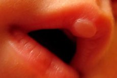
Callus in a newborn baby: on the upper lip, bony
Last reviewed: 08.07.2025

All iLive content is medically reviewed or fact checked to ensure as much factual accuracy as possible.
We have strict sourcing guidelines and only link to reputable media sites, academic research institutions and, whenever possible, medically peer reviewed studies. Note that the numbers in parentheses ([1], [2], etc.) are clickable links to these studies.
If you feel that any of our content is inaccurate, out-of-date, or otherwise questionable, please select it and press Ctrl + Enter.
In pediatrics, a newborn is considered to be a child within four weeks of birth, and during this short time a callus can appear on a newborn: not only on the lip, but also on the bone.
A newborn's lip callus is a sucking pad
Many nursing mothers are concerned about the so-called sucking or milk callus on the lip of a newborn during breastfeeding.
Understanding the reason for its appearance on the baby's upper lip can relieve their anxiety.
Of the more than seven dozen innate reflexes present in newborns, one of the main ones is the sucking reflex, and the main cause of a callus on the upper lip, sometimes in the form of a blister, is repeated vigorous sucking of milk from the breast or from a bottle.
In newborn babies, the oral cavity has some features that help the baby "get" food. Sucking during breastfeeding, as well as when feeding with adapted milk formulas, occurs with the help of jaw and tongue movements. And it begins with the compression of the nipple (or teat) by the baby's lips - due to a strong contraction of the circular muscle of the mouth (musculus orbicularis oris) located in the lips and the movement of the masticatory muscles (musculus masseter) of the lower jaw, which move it in the anteroposterior plane. This compression creates the increased pressure above the nipple necessary for sucking out milk. Then the baby dynamically squeezes milk from the breast into the oral cavity, squeezing the nipple with the tongue towards the hard palate.
At this time, the pressure in the mouth is lower, which is ensured not only by the compression of the lips (the muscle that compresses them, the musculus labii proprius Krause, works), but also by the closure of the internal nasal passages by the soft palate and the lowering of the lower jaw.
In addition, the inner zone of the red border of the upper lip of newborns is larger than the lower one, and has a thicker and higher epithelium with papillae - villous epithelium (under which there is a layer of loose connective tissue). This causes the formation of the pars villosa elevation on the border with the mucous epithelium of the lip, which helps the baby to grasp and hold the nipple.
As neonatologists note, the development of the medial tubercle of the upper lip can occur in the fetus after the 9-10th week of pregnancy (when it begins to suck its thumb in the womb), and in a newborn it has the appearance of a rounded bulge up to 5 mm in size. And this tubercle, although it is a normal anatomical variant, is most often called a callus and only occasionally - a sucking pad. The callus can be permanent, but in some babies it becomes less pronounced 10-15 minutes after the end of each feeding.
True, intense sucking can lead to the formation of a bulla (blister) with serous transparent fluid on this tubercle, and the bubble can burst. But healing occurs spontaneously - without treatment - due to rapid re-epithelialization.
A callus on a newborn's lip does not cause discomfort and does not require treatment: after a few months it disappears on its own.
Bone callus in a newborn is the result of a fracture
It is generally accepted that a bone callus in a newborn appears as a result of birth injuries, primarily a fracture of the clavicle, although fractures of other locations are possible: the humerus and even the femur, during the healing of which new tissue is formed - a bone callus in a newborn.
Risk factors for fractures include: shoulder dystocia during vaginal delivery – difficulty in the midwife’s removal of the shoulder girdle; complicated labor; breech presentation of the fetus (increasing the likelihood of a femur fracture).
Foreign statistics claim that clavicle fractures occur in approximately one newborn out of every 50-60; according to other data, such trauma is observed in no less than 3% of physiological births.
In turn, obstetricians note an increased risk of shoulder dystocia (and clavicle fracture) in cases of high birth weight of the child – fetal macrosomia (≥4500-5000 g); in cases of using vacuum or forceps during childbirth; in gestational diabetes (in diabetic mothers, children have wider shoulders, chest and abdominal circumference); in repeated births – dystocia of the shoulder of the newborn during the first birth (the frequency of recurrent dystocia is estimated at almost 10%).
Therefore, most often, a bone callus forms after a fracture of the clavicle in a newborn.
When considering the pathogenesis of neonatal clavicle fracture, experts emphasize that the process of ossification (ossification) of the tubular clavicle (clavicula) - from the epophyseal plate in its central part - begins in the embryo in the fifth week of intrauterine development. At the same time, the medial part of the clavicle is the thinnest, and the growth plate is open at the time of birth, meaning that the bone is much more susceptible to damage.
In addition, such fractures in newborns are subperiosteal, in which the periosteum is not damaged, and the bones themselves are still soft and often bend in the damaged part without pronounced deformation. Surgeons call fractures of young soft bone green stick fractures. In this case, the formation of subperiosteal new bone and bone callus begins six to ten days after the fracture.
Most often, the symptoms of a fracture are manifested by local swelling, reddening of the skin, hematoma formation, crying of the child when moving the ipsilateral upper limb or lack of movement. This is called pseudoparalysis: the child simply stops moving his arm due to pain.
The consequences and complications of such a fracture develop very rarely: if the area of damage affects the growth plate of the bone (Salter-Harris fractures), and a bridge is formed at the site of the fracture, due to which the growth of the bone is delayed, or it becomes curved.
The diagnosis consists of an examination of the newborn by a pediatrician-neonatologist - with palpation of the clavicles, during which the presence of crunching gives grounds to diagnose a clavicular fracture. The child is also checked for the presence of the Moro reflex, and if it is one-sided (asymmetrical), then the diagnosis of a fracture is confirmed.
In doubtful cases, instrumental diagnostics can be used - ultrasound of the clavicle area. As clinical practice shows, in some cases the damage to the clavicle is so insignificant that it is diagnosed only when the bone callus begins to form in the newborn - with the appearance of a small bulge (bump) on the clavicle, which is a sign of fracture healing.
Differential diagnostics are also carried out: doctors can detect a rare genetic bone disease in a newborn – osteogenesis imperfecta, myotonic dystrophy or multiple joint contractures – arthrogryposis.
What treatment is needed if a newborn has a broken collarbone? Almost all such fractures - due to the great regenerative potential of the periosteum - heal well without therapy as such. But it is necessary to minimize the pressure and movement of the child's arm on the side of the broken collarbone: immobilization is carried out by attaching a sleeve of clothing on the side of the fracture in the front part, while the baby's arm will be bent at the elbow, and the shoulder and forearm are fixed to the body. In case of severe crying, the doctor may prescribe a painkiller, for more details see - Rectal painkillers and anti-inflammatory suppositories.
Normally, a child begins to move the arm on the side of the fracture after about two weeks.
As the researchers found out, the soft callus at the fracture site consists of cartilage and, starting to grow on one side of the fracture, creates a force that aligns the damaged bone. Hardening of the callus promotes complete healing of the fracture, which takes an average of four to five weeks.
Prevention of shoulder dystocia, recommended by some clinicians, involves elective cesarean sections for pregnant women who have a history of delivering a newborn with a clavicle fracture. But experts from the American College of Obstetricians and Gynecologists (ACOG) consider the benefit of this preventive measure questionable.
Additionally, emergency cesarean section carries a higher risk of long bone fracture than normal delivery.
So many experts are inclined to think that it is unlikely that a neonatal clavicle fracture can be prevented during childbirth.
However, the prognosis for a clavicle fracture during childbirth is excellent, and the callus in a newborn after its fracture disappears within six months.