Spring at a newborn baby: when it grows, pathologies
Last reviewed: 17.10.2021

All iLive content is medically reviewed or fact checked to ensure as much factual accuracy as possible.
We have strict sourcing guidelines and only link to reputable media sites, academic research institutions and, whenever possible, medically peer reviewed studies. Note that the numbers in parentheses ([1], [2], etc.) are clickable links to these studies.
If you feel that any of our content is inaccurate, out-of-date, or otherwise questionable, please select it and press Ctrl + Enter.
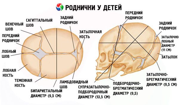
A spring in children is the place where the bones of the skull meet at the site of their supposed fusion. The anatomical features of the structure of the bones of the skull of a newborn child are arranged in such a way that the physiological process of birth can proceed as best as possible. But the changes in the normal appearance and condition of the fontanel in the child can say a lot about the state of his health.
What is fontanel and why is it needed?
Rodnichkom refers to the place on the head of the child, where the bones of the skull do not fuse together and form a connective tissue. Why do we need a fontanel in children, and why the head structure of children is not the same as in adults? The answers are actually very simple. After all, nature has perfectly thought out for the child to undergo gradual changes in the maternal tummy and was born normal and full. When the formation of the bones of the newborn's skull occurs, the processes of osteogenesis are not yet perfect. Therefore, ossicles are soft and pliable in structure. In places where the bones are joined, there should be seams of dense bone tissue, which are represented as fontanel in children. This is because during the delivery, when all the planes of the pelvis pass, the head performs the most important function and regulates the process of passing the child through the birth canal. Therefore, the load and pressure on the skull bones is maximal. Rodnichki allow the bones of the skull to move freely along the generic paths, the bones can be found one on top of the other, which significantly reduces pressure and strain on the brain itself. Therefore, if there were no fontanel in the child, the process of birth would be very complicated.
How many fontanelles does a child have?
A venerated newborn child has only one open fontanel - it's big.
It is located between the frontal bone and two parietal, so it has the irregular shape of the rhombus. If we talk about the total number of fontanelles in a child, then there are six. One front or large, one rear and two lateral on each side. The back fontanel is located between the occipital bone and two parietal. Lateral fontanelles are located on one level - the first between the parietal, temporal and wedge-shaped bone, and the second between the parietal, temporal and occipital. But the lateral fontanelles should be closed at the full term baby, while the front fontanelle is normally opened after the birth of the child and in the first year of his life. Sometimes a full-term child may have a back fontanel, but more often it is closed. The sizes of fontanels in children differ. The largest fontanel is the front font and it is about 25 millimeters in length and width. Next is a small or a back, which is less than 10 millimeters. Lateral fontanelles are the smallest and are no more than five millimeters. To monitor the child's state and the speed of overgrowing of these fontanels, you need to know how to measure the fontanel in a child. This procedure is performed by the doctor every time the child is examined and the result is always recorded on the neonatal development chart. This allows you to monitor the dynamics of the closure of the fontanel. But the mother can also measure at home and this does not require special skills or tools. The large fontanelle has the shape of a diamond, so the measurement is not from corner to corner, but from the side of the diamond to the other side. That is, for measurement, you need to put three fingers of the right hand mom in the projection of the large fontanelle, not in the forward direction in the corners of the rhombus, but slightly in the oblique on the sides of the rhombus. One mom's finger roughly corresponds to one centimeter, and therefore there is no need to measure with a ruler or something else. Thus, the normal size of the fontanel in a child should not exceed the width of three fingers of the mother.
The rates of closure of fontanelles in children differ depending on individual characteristics. After all, one child is breast-fed and has enough minerals and vitamins for early closure of the fontanel, and another child is fed a mixture, and also was born in the winter without a prophylaxis for rickets, therefore, the fontanel is closed at a later date. But still there are normal closing thresholds, the excess of which indicates a possible problem. The great fontanel grows to the 12-18 months of the child's life, and the back or the small one at its opening after birth should close by the end of the second month of the child's life. If the lateral fontanels of a child are open, they should close for six months. When a child grows a fontanel, a dense bone is formed, which will forever be the same as that of an adult.

Pathology of fontanel in children
Naturally, there are certain norms for the closure of fontanelles, but each child may have its own characteristics that affect these terms. Given that the large fontanel is the most revealing and has the most delayed terms of closure, it is always a guideline for the state of health of the infant.
If the fontanelle is closed early in the child, then it is possible to think about metabolic disorders, especially calcium and vitamin D. But it must be remembered that the concept of "early" is very relative, because if the norm is 12 months, and the fontanel was closed at 11 months, then this not so scary. In this case, you should always monitor the dynamics of fontanel sizes throughout the life of the child, because he could have been born with a small fontanel. But if it is a question of closing the big fontanel in 3 months and earlier, then obviously it is necessary to consult with the doctor. This is not always a danger, because you need to assess the overall condition of the baby. Sometimes in young children, the constitutional features of the structure of the head and all parts of the body, in which the children will be small and miniature. Then, for the growth of the brain and head, there is no longer a need for a further increase in the volume of the head, so the fontanel can close earlier. Therefore, it is necessary that the doctor assess the child's condition in a comprehensive manner, taking into account the constitutional features of the development of parents in this period. If we talk about pathology, the early closure of the fontanel in children can be caused by congenital pathologies of the bone system. If there is a pathology of the thyroid gland or parathyroid glands, there may be a fusion of the skull bones on the background of a violation of the level of calcium metabolism. If we talk about congenital malformations, the pathologies of the brain with irregularities in the structure and size of the skull can cause early bone fusion. But if the child was born healthy and normally developed, then moms should not look for him any defect because of the simple early closure of the fontanel.
If the fontanelle is badly overgrown in a child, then there may be more reasons than mom might guess. But in this case, too, it must be remembered that the timing of the growth of the fontanel can be different. If a child has not overgrown with a fontanelle in a year, then this is normal if there is a positive dynamic from birth. For example, if in one month the fontanel was 2.5 by 2.5 centimeters, and in a year it is 1.5 by 1.5 and still does not close, then this is absolutely normal time and by the end of the second half of the child's life it will completely overgrow. But if there is no positive dynamics, then one should think about pathology. The causes of non-overgrowth of the fontanel in a child can be related not only to violations of calcium metabolism, but also there may be other disorders. Rickets can be considered the most common cause of untimely closure of the fontanel. This disease, which is characterized by a deficiency of vitamin D, which violates the absorption and exchange of calcium. This directly affects the state of the bone system of the child, and as a direct sign of pathology the structure of the fontanel is broken. Insufficiency of calcium in the child's body leads to the fact that in the first place there is no normal ossification of the bones of the skull, and the whole process is broken in the child at the place where bone seams should already be formed. This is accompanied by a delay in the closure of the fontanel. Another less common, but more serious problem can be considered congenital hypothyroidism. This disease, which is characterized by a lack of synthesis of thyroid hormones. These hormones in utero and after the birth of a child ensure the active reproduction of all cells and growth of the body. Therefore, the deficiency of these hormones leads to inhibition of active cell growth. Therefore, when the overgrowth of the fontanelle is delayed, along with other symptoms, the pathology of the thyroid gland should be excluded.
If the child has a large fontanel, then this can be a manifestation of hydrocephalus. This is accompanied by an increase in the size of the head against the backdrop of an increase in the volume of its circumference. This pathology develops due to a violation of the outflow of the cerebrospinal fluid through the spinal canal, which is accompanied by the accumulation of this fluid in the brain. But this pathology has a characteristic clinic, which is difficult to miss.
If the child is pulsating the fontanel, and he is tense, then one should think about neurological pathology. Often happens in children who are born in hypoxia or after complicated childbirth after a while the child becomes restless. It begins to pulsate fontanel, especially when it is taken on handles. This may be due to increased intracerebral pressure, which is particularly elevated in the vertical position and causes such a pulsation. But if a child sleeps peacefully, normally eats and does not fuss, then attentive mother sometimes can notice an easy pulsation of the fontanel. This is not an absolute pathology, but it can be a simple pulsation of blood vessels, which is normal for such a baby. Therefore, any pathology of the fontanel is conditional and the doctor's consultation is absolutely necessary.
Sometimes there is a fallen fontanel in a child, which often develops against the background of infection and severe dehydration. The concept of "severe" dehydration for a newborn or an infant is somewhat relative, since even three episodes of diarrhea in such a child can cause symptoms of dehydration. Given that they are of a systemic nature, a decrease in the volume of circulating blood leads to a decrease in the volume of the intracerebral fluid and a decrease in pressure, so the fontanel becomes sunken. This is a very characteristic symptom that can not be ignored.
Often parents are concerned about the tubercle near the fontanel in the child. This may be a simple feature of the fusion of the skull bones, or perhaps a serious neurological pathology. If the tubercle is small and there are no symptoms of anxiety, then it is possible that these are the features of bone adhesion. But if the child is restless or the defect itself is large, then developmental anomalies that require intervention are possible. Therefore, you should always consult a pediatric neurologist.
Rodnichok in premature babies has its own characteristics, since the timing of its overgrowing may be slightly larger. A premature baby can be born with all fontanelles open, depending on the gestation period. It can be strained and pulsating violently due to frequent neurologic symptoms in such babies. In any case, preterm ferns and care for it require special attention.
Rodnikochka in children is the place of the future fusion of the skull bones, which assumes the normal process of the birth of the baby and the further growth of the brain. But although the fontanelle itself consists of connective tissue, its condition can tell about many problems in the child's body. Therefore, it is very important to monitor the condition of the fontanel, the dynamics and timing of its closure, and in time to undergo an examination with a pediatrician.

