New publications
Where to get an ultrasound?
Last reviewed: 02.07.2025

All iLive content is medically reviewed or fact checked to ensure as much factual accuracy as possible.
We have strict sourcing guidelines and only link to reputable media sites, academic research institutions and, whenever possible, medically peer reviewed studies. Note that the numbers in parentheses ([1], [2], etc.) are clickable links to these studies.
If you feel that any of our content is inaccurate, out-of-date, or otherwise questionable, please select it and press Ctrl + Enter.
Where to do an ultrasound, this question has probably arisen for each of us. Let's consider the features of ultrasound examination, the main indications and contraindications for its implementation, as well as the addresses of medical centers and clinics.
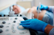
Ultrasound examination is a diagnostic method based on the use of ultrasound waves (air vibrations with a power of 20 kHz to 1000 MHz, inaudible to humans) to examine the subcutaneous structures of the human body. With the help of ultrasound, it is possible to examine tendons, muscles, internal organs, identify disturbances in their functioning and possible pathologies.
The popularity of this diagnostic method is explained by its safety, low cost, and high information content. In addition, ultrasound is one of the few methods that can be repeated if necessary.
There are several modes of operation of ultrasound diagnostic devices, let's consider them:
- One-dimensional mode – the ultrasound beam penetrates into the tissue at one point and is reflected. This mode is used to examine the chambers of the heart, large vessels, cavities, cysts, and wall thickness. The accuracy of this mode and its quality are much higher than in the other two modes.
- Two-dimensional, sectoral or 2D mode – provides a two-dimensional planar image at a certain depth of the located structures. It is the simplest mode for perception, as it reflects the anatomical structure in cross-section.
- Doppler mode – used to evaluate blood flow characteristics and quantitatively evaluate blood flow. Pulse-wave mode allows you to evaluate blood flow at a given depth.
The accuracy of ultrasound examination depends on the diagnostic device, the set of sensors, software and the size of the monitor. The anatomical features of the patient's body, i.e. various pathologies and diseases, also affect the quality of the obtained picture. It is because of these factors that ultrasound can give erroneous information. But there are a number of methods that can increase the accuracy of the study. First of all, the patient must come prepared, i.e. the study of a particular organ requires special preparation. For example, ultrasound of the kidneys is done with a full bladder, and gynecological ultrasound is recommended to be performed in different phases of the menstrual cycle.
But the importance of ultrasound should not be overestimated, since the ultrasound specialist gives a diagnostic conclusion, and the final diagnosis is made only by the attending physician. Therefore, ultrasound data alone is not enough to make a diagnosis; an examination, tests, and a number of other examinations are necessary. In addition, ultrasound is a subjective method, that is, the same data can be interpreted differently by different doctors. Therefore, the qualifications of the doctor and the patient's preparedness for the examination are very important.
Where can I get an ultrasound?
Where can I get an ultrasound and what are the advantages and disadvantages of this method? So, ultrasound diagnostics are performed in medical centers and hospitals equipped with an ultrasound machine and qualified doctors performing the examination.
Let's consider the main types of medical ultrasound in the study of organs and systems of the body:
- Anesthesiology – ultrasound is used when it is necessary to inject an anesthetic into the area around nerve fibers.
- Medical care (emergency) – used to examine the body after injuries. Allows to diagnose disorders of the cardiovascular system and the body.
- Gynecology and obstetrics – identifies bleeding, abnormalities in organ function and other pathologies. Ultrasound is performed on all pregnant women to monitor fetal development.
- Gastroenterology – ultrasound is used to examine abdominal organs. The study reveals inflammatory processes and other pathologies in the pancreas, spleen, stomach and other organs.
- Musculoskeletal system – used to examine ligaments, tendons, bone surfaces, soft tissues, and nerves.
- Cardiology and cardiovascular system – ultrasound examination is used to diagnose heart function and identify abnormalities in the functioning of heart valves and ventricles. With the help of ultrasound, it is possible to determine vascular patency, thrombosis, narrowing of arteries and other pathologies.
- Neurology and ophthalmology – allows us to detect narrowing of the arteries and disturbances in the functioning of nerve fibers.
- Urology – ultrasound is used to examine the pelvic organs and determine disorders in the functioning of the bladder, testicles, and prostate.
As a rule, ultrasound examination is carried out by a doctor's referral. Thus, in some state institutions, ultrasound is done and it is free. But there are many clinics and medical centers that perform ultrasound without a doctor's referral at any time convenient for the patient.
Let's consider the main advantages and disadvantages of ultrasound examination:
Advantages:
- Ultrasound creates live images, which is useful for biopsies, injections, and other imaging procedures.
- The study does not cause discomfort or long-term side effects.
- Allows you to examine muscle and soft tissues, bone surfaces, organs. This is convenient when distinguishing between structures filled with liquid and solid tissues.
- Refers to inexpensive and accessible diagnostic methods, in comparison with computed tomography, magnetic resonance imaging or screening.
Flaws:
- Ultrasound does not provide an image through bone tissue, and when gas forms between the sensor and the organ, the ultrasound is blocked, which negatively affects the accuracy of the study.
- The quality of the image depends on the patient's body type. When diagnosing overweight patients, ultrasound is not always accurate, since the layer of subcutaneous fat blocks the ultrasound of the sensor.
- The accuracy and reliability of the study also depend on the professionalism of the operator, that is, the specialist performing the ultrasound diagnostics.
Where is the best place to get an ultrasound?
Most people do not think about their health and only seek medical help when there are obvious symptoms of illness. You can quickly identify a disease and determine its cause with the help of an ultrasound examination. Where is the best place to do an ultrasound and which clinic to go to, let's consider these issues in more detail.
Ultrasound is based on the ability of tissues of different densities to reflect ultrasound waves. The information received is processed by a special device and an image of organs or tissues is formed on the monitor screen. Ultrasound is used in all areas of medicine and is one of the most common diagnostic methods. Today, many medical centers and hospitals have ultrasound machines at their disposal.
When planning to undergo this examination and choosing the best place to do an ultrasound, you should carefully select the clinic and doctors who will do the diagnostics. This is due to the fact that recently medicine is one of the areas of business, that is, medical ultrasound centers are a kind of way to make money for their owners. But not all of them have a medical education and can correctly interpret the results of the study. Therefore, it is necessary to carefully collect information about the clinics and hospitals where you plan to do an ultrasound.