New publications
It will be easier to diagnose edema
Last reviewed: 02.07.2025

All iLive content is medically reviewed or fact checked to ensure as much factual accuracy as possible.
We have strict sourcing guidelines and only link to reputable media sites, academic research institutions and, whenever possible, medically peer reviewed studies. Note that the numbers in parentheses ([1], [2], etc.) are clickable links to these studies.
If you feel that any of our content is inaccurate, out-of-date, or otherwise questionable, please select it and press Ctrl + Enter.
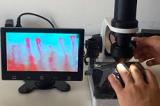
Scientists have developed a new technique for diagnosing edema using the optical wide-field microscopic (capillaroscopic) method and the laser-scanning microscopic method.
The essence of the latest and most sought-after development was described by specialists in the scientific journal Diagnostics.
Until now, practicing physicians have been unable to use quantitative diagnostics and determine the degree of edema, evaluate detailed dynamic changes in edema syndrome. To remedy the current situation, Moscow State University staff have begun developing methods that allow for the precise evaluation of edema characteristics using optical microscopy.
"We were able to demonstrate that for patients with heart failure, the morphological values reflected during vasoscopy can be used to describe long-term edema. Of particular importance here are the diametrical size of the transitional capillary segment and the size of the perivascular section. When examining healthy people, we used two models of short-term edema syndrome and did not reveal any significant changes in capillary values. But in the situations mentioned, we observed a clear decrease in the quality of the image of capillaries, which is due to the accumulation of moisture in the epidermal layer," explained one of the authors of the study.
To verify the obtained results, the scientists used a confocal microscopic method, which provided optimal contrast and intensive spatial expansion during tissue visualization. The work demonstrated that in the presence of edema, papillary-dermal zones acquired hyporefractivity (reduced reflection of optical radiation), which entailed a loss of image contrast. A similar situation was observed in people who underwent infusion therapy. Thus, the proposed diagnostic technique helps to assess the dynamics of edema syndrome in pericapillary zones.
The issue of improving diagnostics for edema has been around for a long time: scientists have been developing new techniques for accessible and reliable clinical examinations of patients for many years. Edematous fluid accumulation in the intercellular space can be observed everywhere in various pathological conditions, including heart failure, inflammatory processes, lymphostasis. The newly developed method will allow practicing doctors to accurately assess the degree and dynamics of edema. Previously, similar diagnostic methods were absent, and doctors had to limit themselves exclusively to a physical examination.