New publications
Optical-acoustic imaging aids in the treatment of spinal muscular atrophy
Last reviewed: 02.07.2025

All iLive content is medically reviewed or fact checked to ensure as much factual accuracy as possible.
We have strict sourcing guidelines and only link to reputable media sites, academic research institutions and, whenever possible, medically peer reviewed studies. Note that the numbers in parentheses ([1], [2], etc.) are clickable links to these studies.
If you feel that any of our content is inaccurate, out-of-date, or otherwise questionable, please select it and press Ctrl + Enter.

Spinal muscular atrophy (SMA) is a severe disease in which a genetic mutation causes degeneration of certain nerves responsible for transmitting signals to muscles. This leads to muscle atrophy, and many patients die a painful death due to this rare disease. Genetic treatments have only become available in the last few years.
Now a team led by Emmanuel Nedoschill, Ferdinand Knieling and Adrian Regensburger from the Translational Pediatrics Working Group in the Department of Pediatrics and Adolescent Medicine at the University Hospital Erlangen has developed a sophisticated procedure that shows encouraging results when used in combination with these treatments: short laser pulses create sound waves that then provide images of muscle tissue.
They published a paper on their findings in the journal Med.
"This method is similar to ultrasound scans that have been around for a long time," explains Nedoshiel. "In a few minutes, a scan from outside the body can provide an image of the condition of the muscles inside the body."
One of the main advantages of this optical-acoustic imaging method is that even small children usually cooperate without much effort, as it is a non-invasive procedure that does not require swallowing or injecting contrast material. This not only simplifies the work of the medical team, but also improves the conditions for children and their parents during their hospital stay.
The situation is usually incredibly stressful for those affected. The disease is caused by just a small change in the genome in terms of a protein called "SNM", but the absence of this protein leads to the degeneration of certain nerves responsible for transmitting signals to muscle cells. The affected muscles atrophy. It can be very difficult for the average person to hear about the consequences and the different ways the disease progresses.
One category is the "ambulators," who are still able to take a few steps on their own. The situation is much worse for the "sedentary." Without help, they can only sit, but cannot stand up on their own. The worst situation is for the "non-sedentary," who cannot even sit. If the muscles needed for swallowing or breathing are affected, the disease can be fatal.
Fortunately, only 1 in about 10,000 newborns has the SNM genetic mutation. However, the suffering of those affected is so great that any improvement in available treatments is a major breakthrough, as is the case with a treatment known as "optoacoustic imaging" (OAI), which is being researched at the Department of Pediatrics and Adolescent Medicine at the University Hospital Erlangen.
These treatments, which became available only a few years ago, have led to significant breakthroughs in the treatment of what was previously a virtually incurable condition. Notable improvements have been achieved even in the most severe cases, called "non-sedentary."
Until now, however, the only way to track this progress has been through grueling motor tests that can last for days. The very nature of these tests can also compromise their objectivity. Some people may exert more effort than others, leading to better results in some children than others. Children’s moods can also vary from day to day, affecting test results.
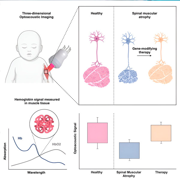
The OAI procedure with short laser pulses using near infrared light can significantly improve the objectivity of these observations. These light pulses heat the affected tissue, which then emits sound waves that provide important information about various structures in the patient’s body. The tissue, for example, is made up of collagen proteins, which return a different spectrum of sound waves compared to muscle or fat tissue.
Source: Med (2024). DOI: 10.1016/j.medj.2024.02.010
"In muscle, we can identify a spectrum of hemoglobin in red blood cells, which is responsible for transporting oxygen to the body and removing carbon dioxide," explains Nedoshill. The more muscle cells there are and the more active they are, the more oxygen they require to do their job.
If a researcher at the University Hospital Erlangen sees a lot of hemoglobin, he knows that this means significant muscle mass. On the other hand, if the muscles atrophy and are replaced by connective tissue, 3D images show how the disease progresses and leads to an increase in collagen, documenting the atrophy of muscle mass.
This gives doctors like Nedoshiel a tool that is as quick and easy to use as an ultrasound scan, and provides dramatic images of how muscles and connective tissue come and go.
Research conducted in Erlangen using haemoglobin tracking has shown that children with SMA have significantly less muscle tissue than healthy controls. However, after receiving life-saving genetic therapy, the haemoglobin concentration increases, atrophied muscles begin to regenerate, and ultrasound signals soon begin to resemble those of healthy organisms.
Thanks to research at the Department of Paediatrics and Adolescent Medicine in Erlangen, a relatively simple tool is now available to monitor the progress of muscle atrophy and the success of treatment.