New publications
Clarification of the cellular mechanisms of periodontitis with an improved animal model
Last reviewed: 02.07.2025

All iLive content is medically reviewed or fact checked to ensure as much factual accuracy as possible.
We have strict sourcing guidelines and only link to reputable media sites, academic research institutions and, whenever possible, medically peer reviewed studies. Note that the numbers in parentheses ([1], [2], etc.) are clickable links to these studies.
If you feel that any of our content is inaccurate, out-of-date, or otherwise questionable, please select it and press Ctrl + Enter.
Researchers from Tokyo Medical and Dental University (TMDU) have developed a technique that allows detailed analysis of the development of periodontitis over time.
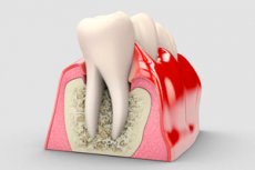
Periodontal disease, known as periodontitis, is the leading cause of tooth loss and affects nearly one in five adults worldwide. In most cases, the condition results from an inflammatory response to a bacterial infection of the tissues around the teeth.
As the condition worsens, the gums begin to recede, exposing the roots of the teeth and bone. Notably, the incidence of periodontitis increases with age, and with the world population living longer, it is important to have a solid understanding of its underlying causes and progression.
In a study published in the journal Nature Communications, researchers from TMDU found a way to achieve this goal by improving a widely used animal model for studying periodontitis.
Directly studying periodontitis in humans is difficult. As a result, scientists often turn to animal models for preclinical studies. For example, the "ligature-induced periodontitis mouse model" has allowed researchers to study the cellular mechanisms underlying the condition since its introduction in 2012.
In simple terms, this model artificially induces periodontal disease by placing silk threads on the molars of mice, causing plaque to accumulate. Although this method is convenient and effective, it does not capture the full picture of periodontitis.

Schematic illustration of inflammatory gene expression profiles during periodontitis and the role of the IL-33/ST2 axis in combating acute inflammation. Source: Tokyo Medical and Dental University.
"Although periodontal tissue consists of gingiva, periodontal ligament, alveolar bone, and cementum, analysis is typically performed solely on gingiva samples due to technical and quantitative limitations," notes lead study author Anhao Liu. "This sampling strategy limits the conclusions that can be drawn from these studies, so methods that allow for simultaneous analysis of all tissue components are needed."
To address this limitation, the research team developed a modified ligature-induced periodontitis model. Instead of the classic single ligature, they used a triple ligature on the upper left molar of male mice. This strategy expanded the area of bone loss without significantly destroying the bone around the second molar, increasing the number of different types of periodontal tissue.
"We isolated three main tissue types and assessed the RNA yield between the two models. The results showed that the triple-ligated model effectively increased the yield, achieving four times the amount of normal periradicular tissue and supporting high-resolution analysis of different tissue types," explains senior author Dr. Mikihito Hayashi.
After confirming the effectiveness of their modified model, the researchers set out to study the effects of periodontitis on gene expression across different tissue types over time, focusing on genes associated with inflammation and osteoclast differentiation.
One of their key findings was that the expression of the Il1rl1 gene was significantly higher in the periradicular tissue five days after ligation. This gene encodes the ST2 protein in receptor and decoy isoforms, which binds to a cytokine called IL-33, which is involved in inflammatory and immunoregulatory processes.
To gain further insight into the role of this gene, the team induced periodontitis in genetically modified mice that lacked either the Il1rl1 or Il33 genes. These mice showed accelerated inflammatory bone destruction, highlighting the protective role of the IL-33/ST2 pathway. Further analysis of cells containing the ST2 protein in its receptor form, mST2, revealed that most of them originated from macrophages.
"Macrophages are generally classified into two main types, pro-inflammatory and anti-inflammatory, depending on their activation. We found that mST2-expressing cells were unique in that they simultaneously expressed some markers of both types of macrophages," commented senior author Dr. Takanori Iwata. "These cells were present in the periradicular tissue before inflammation began, so we named them 'periodontal tissue-resident macrophages.'"
Together, the results of this study demonstrate the power of a modified animal model to study periodontitis at a more detailed scale, down to the biomolecular level.
"We propose the possibility of a novel IL-33/ST2 molecular pathway regulating inflammation and bone destruction in periodontal disease, along with specific macrophages in the periradicular tissue that are deeply involved in periodontal disease. This will hopefully lead to the development of new treatment strategies and prevention methods," concludes senior author Dr. Tomoki Nakashima.