New publications
Miniature optical coherence tomography probe takes images inside cerebral arteries
Last reviewed: 02.07.2025

All iLive content is medically reviewed or fact checked to ensure as much factual accuracy as possible.
We have strict sourcing guidelines and only link to reputable media sites, academic research institutions and, whenever possible, medically peer reviewed studies. Note that the numbers in parentheses ([1], [2], etc.) are clickable links to these studies.
If you feel that any of our content is inaccurate, out-of-date, or otherwise questionable, please select it and press Ctrl + Enter.
An international team of microtechnologists, medical technologists and neurosurgeons have designed, built and tested a new type of probe that can be used to take images from inside the brain's arteries.
In their paper published in the journal Science Translational Medicine, the group describes how the probe was designed and built, as well as how it performed in initial tests.
When patients have medical problems in the brain, such as blood clots, aneurysms, or hardened arteries, the tools available to doctors to diagnose them are limited to imaging technologies that take pictures of veins and arteries from the outside of the brain. These images are then used as maps to guide catheter-like devices through the veins and arteries into parts of the brain to perform repairs.
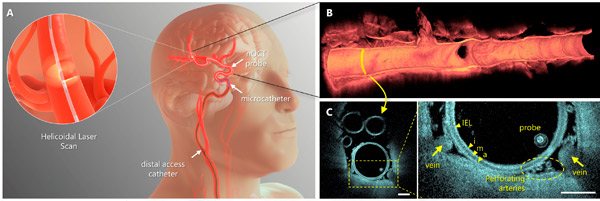
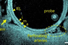
Intravascular imaging using neuro-optical coherence tomography (nOCT). The nOCT probe is compatible with standard neurovascular microcatheters, integrating with the procedural workflow used in clinical practice. nOCT captures high-resolution 3D optical data, providing volume microscopy of tortuous cerebral arteries, surrounding structures, and therapeutic devices. Source: Science Translational Medicine (2024). DOI: 10.1126/scitranslmed.adl4497
The problem with this approach is that the images used are not always clear or accurate. They also do not allow the surgeon to see what is happening inside the vein or artery as it is being repaired, resulting in procedures being performed almost blindly.
In this new study, the team created a camera probe that is small enough to fit inside a catheter, allowing it to capture near-real-time images from inside the brain's veins and arteries.
The new probe is based on optical coherence tomography, a type of imaging technology used by eye and heart surgeons to treat patients. It generates images by processing the backscatter of near-infrared light. Until now, such devices have been too bulky and rigid to be used inside the brain.
To overcome this problem, the research team replaced the components with smaller parts, such as a fiber optic cable as thin as a human hair. They also used a modified type of glass to make the distal lens, which makes up the head of the probe and allows it to bend.
The resulting probe is mostly hollow and worm-shaped. It also rotates at 250 times per second, which helps it move easily through the veins and arteries. The camera takes pictures at a rate proportional to the need. The entire probe fits easily inside the catheter, making it easy to place and move around the arteries and veins of the brain, as well as remove it.
After animal testing, the probe was moved into clinical trials at two sites, one in Canada and one in Argentina. To date, 32 patients have been treated with the new probe. The team reports that so far, the probe has proven safe, well-tolerated, and successful in all cases. They conclude that their new probe is ready for general use.