New publications
Midkine vs. Amyloid: Brain Development Protein Surprisingly Inhibits Aβ Assembly and Plaque Formation
Last reviewed: 23.08.2025

All iLive content is medically reviewed or fact checked to ensure as much factual accuracy as possible.
We have strict sourcing guidelines and only link to reputable media sites, academic research institutions and, whenever possible, medically peer reviewed studies. Note that the numbers in parentheses ([1], [2], etc.) are clickable links to these studies.
If you feel that any of our content is inaccurate, out-of-date, or otherwise questionable, please select it and press Ctrl + Enter.

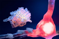
In the giant proteomic catalogs of the Alzheimer's brain, one underappreciated player keeps popping up: midkine (MDK). This protein is markedly elevated early in the disease and closely correlates with amyloid-β (Aβ), but its role in the pathology has long remained a mystery. The St. Jude team and partners went "molecule to animal model" and showed that MDK weakens the assembly of Aβ fibrils and affects the formation of amyloid plaques. In essence, it is a natural "antiplatelet" of Aβ, which the brain itself increases in the disease.
Background of the study
Alzheimer's disease is currently treated in the "anti-amyloid paradigm": antibodies to amyloid-β (Aβ) do clear plaques and moderately slow cognitive decline in the early stages. In 2023, the FDA approved lecademab, in 2024 - donanemab; in parallel, there are debates about the balance of benefits and risks (ARIA-edema/hemorrhage), availability and cost, as can be seen from the decisions of EMA/NICE and discussions in the clinical press. The therapeutic picture is improving, but remains "narrow": additional targets and approaches are needed that not only remove already formed plaques, but also prevent Aβ aggregates themselves from arising and growing.
One of the promising ways is to rely on the endogenous antiplatelet mechanisms of the brain. People have been described to have their own proteins, "chaperones", which in vitro and in models can interfere with the early stages of Aβ assembly: clusterin, apolipoprotein E, transthyretin, the BRICHOS domain, etc. The picture is ambiguous: some proteins in physiological concentrations delay the start of fibrillogenesis, while others in certain contexts, on the contrary, can promote fibrillation or cellular capture of "seeds" - hence the interest in those endogenous moderators whose role in Aβ is stable and reproducible.
Against this background, attention was drawn to midkine (MDK), a heparin-binding growth factor known for its roles in the development of the nervous system, regeneration, and inflammation. In proteomic sections of the brain in Alzheimer's, MDK is consistently elevated already in the early stages and correlates with Aβ, but for a long time it remained unclear whether it was simply a "marker of trouble" or an active participant in the process. The biology of midkine suggests both possibilities: it is a stress-induced protein that changes with a wide variety of damage both in the central nervous system and on the periphery, interacting with several receptor systems.
A new paper in Nature Structural & Molecular Biology closes this “knowledge gap” by moving from observation to mechanistic: it shows that MDK physically binds to Aβ and inhibits fibrillogenesis in a multi-angle panel of methods (ThT, CD, EM, NMR), and in the 5xFAD model, knocking out Mdk increases amyloid burden and microglial activation. In other words, the brain itself seems to raise a “natural antiplatelet,” and its loss exacerbates pathology – a thesis that makes MDK an attractive axis for both risk/progression biomarkers and therapeutic mimetics capable of supporting endogenous defense alongside antibodies.
How they tested: from test tubes and spectra to transgenic mice
First, the researchers looked into the chemistry: how recombinant MDK affects Aβ40 and Aβ42 fibrillogenesis. To do this, they conducted fluorescence tests with thioflavin T, circular dichroism, negative contrast electron microscopy, and NMR in parallel. All the methods agreed: MDK inhibits fibril formation and binds to Aβ threads isolated from human AD brain. Then came the physiology: in the 5xFAD amyloidosis model, genetic knockout of Mdk led to greater Aβ accumulation, increased microglial activation, and plaque growth; on the contrary, the presence of midkine “kept” the pathology lower. Finally, mass spectrometric proteomic analysis (complete and detergent-insoluble proteome) confirmed that in the absence of Mdk, Aβ and associated protein networks, as well as microglial components, grow in the mouse brain. Together, this adds up to a picture of a protective role for MDK against amyloid pathology.
What exactly did they do and measure?
- In vitro: Aβ40/Aβ42 + MDK → ThT fluorescence, CD, negative CEM, and NMR “rescue” of Aβ monomer signals, which are usually “silenced” by aggregation.
- Ex vivo/in situ demonstration of MDK association with Aβ filaments from AD patient brains.
- In vivo: Mdk knockout in the presence of 5xFAD → more plaques and microglial activation; further - proteomics of whole tissue and the "insoluble" fraction, where aggregates accumulate.
- Open data: NMR shifts have been uploaded to BMRB 17795, raw proteomic files have been uploaded to PRIDE (PXD046539, PXD061103, PXD045746, PXD061104).
Key findings
The key result is that midkine prevents Aβ from assembling into stable fibrils, and its absence in the living brain aggravates amyloid pathology. Midkine colocalizes with Aβ in human samples and physically interacts with the filaments, which is consistent with the idea of a "natural brake" on aggregation. In mice without Mdk, not only Aβ itself grows, but also the "accompanying" proteins of its network and signs of microglial activity - a sure indicator of an increase in the inflammatory component of the pathology.
Why is this important in the context of the “anti-amyloid era”
We have entered the era of anti-Aβ antibodies, but they are far from a “silver bullet”: moderate efficacy, the risk of ARIA, and strict selection criteria limit their use. The emergence of an endogenous fibrillogenesis moderator opens an alternative path: supporting the brain’s own antiplatelet mechanisms. There are many options, from MDK domain mimetics and stabilizing compounds to biological strategies for increasing its activity in the right compartments. But before talking about therapy, rigorous testing of safety and long-term effect in large animals and in humans is needed.
How this can be useful already at the research stage
- Biomarker axis: MDK level/localization as a stratification marker of the risk of rapid increase in amyloid load (in conjunction with PET-Aβ and cerebrospinal fluid parameters).
- Combined approaches: “soft” antiplatelet background via the MDK pathway + targeted elimination of existing Aβ (antibody) can theoretically provide additivity.
- Structural clues: NMR/CEM data will suggest MDK-Aβ interaction sites for small molecule/peptide design.
How methods “see” it: a bit of technique
Spectroscopic triangulation is important because each method captures a different aspect of aggregation: ThT is sensitive to fibril β-sheets; circular dichroism tracks conformational transitions; CEM shows filament morphology; NMR captures the “disappearance” of monomer signals as complexes become larger. Here, MDK reduced the ThT signal, shifted the CD spectra, changed the CEM filament pattern, and returned the Aβ NMR signals, consistent with slowing and/or rerouting the aggregation pathway. In 5xFAD brains without Mdk, the picture is mirrored: more Aβ and satellite proteins, plus microglia “on edge.”
Important limitations - do not confuse "effect" with "medicine"
This is fundamental work: test tube + mice. It shows a role for MDK in amyloid biology, but does not prove that increasing midkine is safe and beneficial for long-term therapy in humans. MDK has a broad biology (development, regeneration, inflammation), so systemic interventions may have ambiguous consequences; the true “dose-target-compartment” in the brain remains an open question. Finally, 5xFAD is a powerful but particular model of amyloid pathology; confirmation in other models and in humans is needed for clinical relevance.
What is the logical thing to do next?
- To map MDK-Aβ interaction domains and test mimetics/anti-aggregation peptides in vivo.
- To test the dose-response and safety of long-term elevation of MDK in the brain of large animals.
- To compare CSF/plasma MDK levels with PET-Aβ dynamics and cognitive trajectories in humans (longitudinal cohorts).
Briefly - three facts
- Midkine (MDK) is an endogenous protein that attenuates Aβ40/Aβ42 fibrillogenesis and is associated with amyloid filaments from AD brain.
- Knockout of Mdk in the 5xFAD model leads to more plaques, accumulation of Aβ-related proteins and microglial activation.
- This is a candidate defense axis that can be developed as a biomarker and therapeutic direction, but there are still several stages of testing before it reaches the clinic.
Source: Zaman M. et al. Midkine attenuates amyloid-βfibril assembly and plaque formation. Nature Structural & Molecular Biology, August 21, 2025. DOI: https://doi.org/10.1038/s41594-025-01657-8
