New publications
Maternal microbiota programs development of stress node in future offspring
Last reviewed: 18.08.2025

All iLive content is medically reviewed or fact checked to ensure as much factual accuracy as possible.
We have strict sourcing guidelines and only link to reputable media sites, academic research institutions and, whenever possible, medically peer reviewed studies. Note that the numbers in parentheses ([1], [2], etc.) are clickable links to these studies.
If you feel that any of our content is inaccurate, out-of-date, or otherwise questionable, please select it and press Ctrl + Enter.

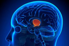
A paper published in Hormones and Behavior shows that the gut microbiota sets the parameters for the development of the paraventricular nucleus of the hypothalamus (PVN), a key center for the stress response. Mice raised without microbes (germ-free, GF) had fewer cells in the PVN both in the neonatal period and in adulthood, without changing the volume of the nucleus (i.e., it is the cell density that decreases). Cross-feeding showed that the effect is programmed even before birth, through the maternal microbiota.
Background
What is the PVN and why is it important?
The paraventricular nucleus of the hypothalamus (PVN) is a “hub” of the stress system: its CRH neurons trigger the hypothalamic-pituitary-adrenal (HPA) axis and influence behavior, motivation, water-salt balance, and energy metabolism. Therefore, any shifts in the cellular composition of the PVN potentially alter stress reactivity and homeostasis.
Microbiota and the Stress Axis: Classic Data
Even in “classical” experiments, it was shown that in mice raised without germs (germ-free, GF), the HPA axis stress response is hyperreactive; colonization with “friendly” bacteria (e.g., Bifidobacterium) partially normalizes this phenotype. This was the first direct clue that intestinal microbes “tune” the stress neuroendocrine system.
Maternal Microbiota and Prenatal Brain Development
It was later discovered that the effect begins before birth: depletion of the microbiota in pregnant females (antibiotics/GF) disrupts the expression of axonogenesis genes in the embryo and the formation of thalamocortical pathways; likely mediators are microbially modulated metabolites that signal to the developing brain. This has been documented in Nature -level papers.
Neuroimmune “gearbox”: microglia
Gut microbes drive the maturation and function of microglia, the master gardeners of the developing brain that regulate apoptosis/synaptic pruning and inflammatory responses. In the absence of microbiota, microglia are immature and functionally defective; restoration of the microbial community partially rescues the phenotype. This provides a mechanism by which peripheral microbiota can rewire neuronal circuits.
Why the focus on the PVN now?
The PVN is the apex of the HPA and is also a node sensitive to early stressors and nutritional cues. Evidence has emerged that PVN^CRH neuron activity not only drives the cortisol response but also influences behavior/motivation; therefore, changes in PVN cellular architecture may have long-term consequences for stress resilience.
What was missing before the current work
It was known that (a) the microbiota “spins” the HPA axis and (b) the maternal microbiota programs neurodevelopmental trajectories. But there was a gap: is there an anatomical trace of this specifically in the PVN — does the number/density of cells change and when is the “sensitivity window” open (before or after birth)? The work in Hormones and Behavior closes this gap: in the absence of microbiota, mice have a decrease in the number of PVN cells in newborns and adults without changing the volume of the nucleus, and cross-feeding shows that programming begins prenatally.
Implications and the Next Mile
If the maternal microbiota sets the PVN cell density in utero, then microbiota modifiers (maternal diet, antibiotics, infections, probiotics/postbiotics) may influence the “tuning” of the stress axis in the offspring. Further work will require: single-cell PVN profiles (which neurons - CRH/AVP/OT - are affected), tests of HPA function and behavioral phenotypes in adults, and testing the role of specific metabolites (e.g., short-chain fatty acids) as signaling molecules between the gut and the developing brain.
How was this tested?
The authors compared the offspring of normal (colonized) mice (CC) and sterile (GF) mice, and also used cross-feeding immediately after birth:
- CC → CC (control),
- GF → GF (sterile mothers and sterile pups),
- GF → CC (sterile pups transplanted to normal mothers).
On the 7th day of life, the GF → GF and GF → CC mice had a lower cell count in the PVN than the CC → CC mice, with the PVN volume remaining the same — hence the decrease in cell density. The second experiment in adult GF mice also confirmed a decrease in the cell count in the PVN (with the volume remaining the same). There are two conclusions: 1) increased cell death in GF newborns leaves a permanent mark; 2) since transplantation to “microbial” mothers on the day of birth did not correct the deficiency, the maternal microbiota sets the developmental trajectory already in the womb. It was additionally noted that the microbiota status and gender affect the overall forebrain size (larger in GF mice; larger in females), without any interaction of the factors.
Why is this important?
The PVN is a nodal structure that initiates the stress response axis (HPA), and is involved in the regulation of autonomic functions, water-salt balance, and nutrition. If the maternal microbiota “twists” the number of neurons in the PVN before birth, this adds a direct anatomical link to the growing “microbiota-brain” chain and helps explain why early factors (nutrition, antibiotics, childbirth) have such a significant impact on stress resistance and behavior later in life. The result fits logically with previous observations on the influence of the microbiota on perinatal neuronal and microglia death.
What this does not prove (limitations)
- This is a mouse model: transfer to humans requires caution.
- The change in "cell number" does not directly indicate which neurons are affected (e.g. CRH neurons of the PVN) or how the function changes (stress hormones, behavior).
- The mechanism remains open: are these microbial metabolites (short-chain fatty acids, etc.), immune signals, or interactions with glia? Targeted experiments are needed. (Reviewed literature points to both pathways.)
What's next?
- Single-cell PVN transcriptomes after microbiota manipulations (including selective metabolite rescues) and functional assays of the HPA axis.
- Testing to what extent the “window of sensitivity” is limited to the intrauterine period and early postnatal time.
- The relationship between anatomical changes and behavioral phenotypes in adults (stress reactivity, nutrition, sleep) - and whether they can be “fixed” later.
Source: Hormones and Behavior, Epub 21 Apr 2025; Print Jun 2025 (Vol 172, Article 105742). Authors: YC Milligan et al., Georgia State University Neuroscience Institute. https://doi.org/10.1016/j.yhbeh.2025.105742
