New publications
The largest 3D reconstruction of a fragment of the human brain has been created
Last reviewed: 02.07.2025

All iLive content is medically reviewed or fact checked to ensure as much factual accuracy as possible.
We have strict sourcing guidelines and only link to reputable media sites, academic research institutions and, whenever possible, medically peer reviewed studies. Note that the numbers in parentheses ([1], [2], etc.) are clickable links to these studies.
If you feel that any of our content is inaccurate, out-of-date, or otherwise questionable, please select it and press Ctrl + Enter.
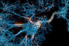
A cubic millimeter of brain tissue may not seem like much. But when you consider that that small square contains 57,000 cells, 230 millimeters of blood vessels, and 150 million synapses, totaling 1,400 terabytes of data, the Harvard and Google researchers have made huge progress.
A Harvard team led by Jeff Lichtman, the Jeremy R. Knowles Professor of Molecular and Cellular Biology and newly appointed dean of the Faculty of Science, and Google researchers have created the largest 3-D reconstruction of the human brain to date at the synaptic level, showing in vivid detail every cell and its network of neural connections in a portion of the human temporal cortex about the size of half a grain of rice.
The achievement, published in the journal Science, is the latest in a nearly decade-long collaboration with scientists at Google Research that combines Lichtman electron microscopy with AI algorithms to color-code and reconstruct the highly complex neural wiring of mammals. The three co-first authors of the paper are former Harvard postdoc Alexander Shapson-Ko, Google Research’s Michal Januszewski, and Harvard postdoc Daniel Berger.
The ultimate goal of the collaboration, supported by the National Institutes of Health's BRAIN Initiative, is to create a high-resolution map of neural connectivity across the entire mouse brain, which would require about 1,000 times more data than they just got from one cubic millimeter of human cortex.
The word "fragment" is ironic. A terabyte is a huge amount to most people, but a fragment of a human brain - just a tiny, tiny part of a human brain - is still thousands of terabytes."
Jeff Lichtman, Jeremy R. Knowles Professor of Molecular and Cellular Biology
The latest map in Science contains details of the brain’s structure that have not been seen before, including a sparse but powerful network of axons connected by up to 50 synapses. The team also noted some peculiarities in the tissue, such as a small number of axons forming extensive spirals. Because their sample was taken from a patient with epilepsy, they are unsure whether such unusual formations are pathological or simply rare.
Lichtman's area of research is "connectomics," which, like genomics, seeks to create complete catalogs of brain structure down to individual cells and connections. Such complete maps will illuminate the path to new understandings of brain function and diseases about which scientists still know very little.
Google's state-of-the-art AI algorithms can reconstruct and map brain tissue in three dimensions. The team has also developed a set of publicly available tools that researchers can use to explore and annotate the connectome.
"Given the huge investment that went into this project, it was important to present the results in a way that someone else could now benefit from them," said Google Research fellow Viren Jain.
Next, the team will target a region of the mouse hippocampus that is important to neuroscience because of its role in memory and neurological disease.