New publications
The world's first atlas of the human brain has been created
Last reviewed: 01.07.2025

All iLive content is medically reviewed or fact checked to ensure as much factual accuracy as possible.
We have strict sourcing guidelines and only link to reputable media sites, academic research institutions and, whenever possible, medically peer reviewed studies. Note that the numbers in parentheses ([1], [2], etc.) are clickable links to these studies.
If you feel that any of our content is inaccurate, out-of-date, or otherwise questionable, please select it and press Ctrl + Enter.
A European team of scientists, which included 12 groups of specialists from Germany, England, Israel, Switzerland, Italy, France and Denmark, completed work on creating an atlas of the human brain – the most detailed analysis of the microstructure of the human brain to date. In addition, the specialists made a cartography of the white matter of the human brain.

The project took three years to complete and required considerable investment, namely 2.5 million euros, and finally the experts presented the fruits of their labor.
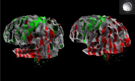
The neuroanatomical picture of the brain was compiled based on the analysis of brain activity processes of 100 volunteers. The brain was examined using magnetic resonance scanning.
To obtain a three-dimensional image of bundles of neurons inside the brain, the researchers used a technique called diffusion tensor imaging.
"The human brain is the most complex structure known to man, and also the most difficult puzzle for modern science, which seeks to understand how it is organized and structured. Our study provides the first opportunity to get closer to understanding the connection between the brain and the genome, and also indicates that genetic disorders can cause brain diseases," say the authors of the study.
Currently, a brain atlas is being used worldwide, which was compiled thanks to two volunteers who donated their bodies for the benefit of science and new discoveries.
The new atlas is innovative in that it shows microscopic features in the white matter, which contains the neural fibers that transmit information throughout the brain.
The project's results, obtained using advanced imaging techniques, provide new depth and precision in understanding human brain processes both in health and in disease.
These images will serve as a benchmark for future brain research and will also be used for medical purposes.
The findings will greatly aid future research into the brain's white matter. Historically, the vast majority of research has focused on understanding and studying gray matter and neurons, while white matter has received relatively little attention.
With the help of the new atlas, investigators and doctors will be able to compare samples with images of the corresponding structures of the brain of a healthy person. The brain atlas is undoubtedly a very useful thing in the development of new diagnostic methods, and is also important in studying the processes occurring in the human brain.

 [
[