
New publications
Cells with giant nuclei may mark the earliest sites of cancer formation.
Last updated: 15.09.2026

All iLive content is medically reviewed or fact checked to ensure as much factual accuracy as possible.
We have strict sourcing guidelines and only link to reputable media sites, academic research institutions and, whenever possible, medically peer reviewed studies. Note that the numbers in parentheses ([1], [2], etc.) are clickable links to these studies.
If you feel that any of our content is inaccurate, out-of-date, or otherwise questionable, please select it and press Ctrl + Enter.
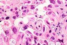
Researchers from Nagoya University have discovered a cellular condition that can arise in the earliest stages of tumor formation—even before full-blown cancer develops. Chronic oxidative damage from iron killed some renal tubule cells, but some survived, developed extremely large and dense nuclei, and simultaneously activated programs associated with resistance to cell death and tumor transformation. The study was published in the journal Redox Biology.
These cells are called karyomegalic—their nuclei are significantly larger than normal. Pathologists have observed similar changes before, but it remained unclear whether they were simply a consequence of severe tissue damage or whether they represented cells that could later develop into tumors. The new study shows that, at least in an experimental model, some karyomegalic cells acquire a characteristic set of molecular features that enable them to survive dangerous oxidative stress.
This process was particularly pronounced in animals with partial BRCA1 gene deficiency. These animals exhibited an increased number of karyomegalic cells with increased activity of the MYC, MET, and LCN2 genes, altered iron metabolism, and impaired mitochondrial function. At the same time, surrounding cells also began to acquire similar characteristics, creating not just a single abnormal cell, but an entire localized precancerous environment.
Most interestingly, the molecular programs identified in animals had analogs in human kidney cancer data. In the Cancer Genome Atlas database, the activity of the most unfavorable cellular profile was associated with worse clinical outcomes, while another variant of karyomegaly, which retained signs of normal metabolism, was associated with a more favorable prognosis. Furthermore, in a small analysis of breast tissue, similar nuclear shape abnormalities were more frequently observed in carriers of inherited BRCA1 mutations.
| The main result | What was discovered? |
|---|---|
| Chronic oxidative stress | Causes severe damage to the renal tubular epithelium |
| Some cells do not die. | Cells with abnormally large nuclei are formed |
| Karyomegaly | Associated with the restructuring of gene activity and metabolism |
| BRCA1 deficiency | Enhances the emergence of a potentially dangerous cellular condition |
| Ferroptosis | Precancerous cells acquire signs of resistance to this type of death. |
| Mitochondria | Their structure and energy function are disrupted. |
| Cell environment | Neighboring cells also enter a stress-resistant state. |
| People's data | Similar gene programs are associated with prognosis in kidney cancer. |
How scientists modeled the early stages of tumor formation
The researchers used an experimental model of kidney carcinogenesis in rats based on exposure to iron nitrilotriacetate. This compound induces reactions involving iron and the formation of highly reactive free radicals that damage lipids, proteins, and DNA. This model is used to study how prolonged oxidative stress can gradually transform normal renal tubular epithelium into malignant tissue.
Scientists compared normal animals and rats with only one full copy of Brca1 due to the L63X mutation. This condition is called haploinsufficiency: the gene is not completely silenced, but the amount of its normal product is reduced. BRCA1 is particularly known as a protein involved in DNA damage repair and tumor suppression, so the researchers were interested in whether its partial deficiency would alter the cells' ability to survive oxidative damage.
Six kidney samples were used for spatial analysis: control tissue, tissue after one week of iron exposure, and tissue after three weeks of exposure—separately for normal and Brca1-mutant animals. Xenium spatial transcriptomics was used on each sample, allowing for the determination of gene activity in individual cells directly within the preserved tissue. The specialized panel included 50 genes associated with kidney structure, mitochondria, iron metabolism, ferroptosis, and carcinogenesis.
The volume of spatial data obtained was enormous: in each sample, the algorithm identified approximately 325,000 to 531,000 cells. After RNA analysis, the same sections were stained with hematoxylin and eosin, allowing the scientists to directly correlate the shape of a specific nucleus with the gene activity of that particular cell and its location within the kidney tissue.
This is a significant advantage of the study. Conventional molecular tissue analysis reveals the average gene activity of thousands or millions of cells, but it doesn't allow us to understand which cells have changed or who is nearby. Spatial transcriptomics allowed the researchers to simultaneously observe the cellular morphology, molecular state, and microenvironment surrounding a potentially precancerous cell.
| Method | What it allowed to determine |
|---|---|
| Iron damage model | Create chronic oxidative stress in the kidney |
| Brca1 mutant rats | To study the influence of hereditary predisposition |
| Spatial transcriptomics | Determine gene activity of individual cells in situ |
| A panel of 50 genes | Assess iron metabolism, mitochondria, ferroptosis, and tumor signaling |
| Histological staining | See the shape and size of the nucleus |
| Digital morphometry | Quantitatively measure the characteristics of the core |
| Spatial analysis | Determine the state of adjacent cells and stroma |
After exposure to iron, normal cells began to lose their specialization
The first changes were noticeable after just one week of exposure. In normal proximal renal tubule cells, genes required for substance transport and energy metabolism, including LRP2, PINK1, and AMACR, are typically active. After oxidative damage, the activity of these normal programs decreased, and genes characteristic of tissue damage, repair, and potential tumor transformation were activated in their place.
Among the most significantly upregulated genes were MYC, MET, LCN2, and ANXA2. MYC is one of the best-known regulators of cell growth and division, and disturbances in its regulation are often found in cancer. MET encodes a receptor capable of stimulating cell growth, survival, and migration. LCN2 is involved in, among other things, iron metabolism and the cellular stress response.
After three weeks, the differences between normal animals and those with BRCA1 deficiency became more pronounced. In normal rats, some cells showed signs of restoration to normal, while in Brca1-mutant kidneys, damaged cellular states persisted and expanded. The activity of normal kidney genes was particularly noticeably reduced, while the activity of tumor and stress-related programs increased.
The researchers identified 11 molecular cell states in the proximal tubules. These included relatively normal cells, cells with antioxidant adaptation, cells with an activated DNA damage response, transiently regenerating cells, cells with an unsuccessful repair program, and cells undergoing ferroptosis. Animals with BRCA1 deficiency showed a particularly elevated proportion of states associated with inadequate tissue repair.
Thus, early carcinogenesis in this model appeared not as the instantaneous appearance of a single tumor cell, but as a gradual restructuring of an entire cellular community. Normal specialized cells lost some of their functions, some died, others recovered, and still others entered a stable stress state, potentially creating conditions for further tumorigenesis.
| Cell state | Characteristic changes |
|---|---|
| Normal tubule cell | High activity of LRP2, PINK1, AMACR |
| Oxidative adaptation | Strengthening antioxidant mechanisms |
| DNA damage | Activating recovery programs |
| Regeneration | Temporary increase in cell division |
| Unsuccessful recovery | Increased LCN2 and other stress signals |
| Ferroptosis | Iron-dependent death of damaged cells |
| Precancerous condition | Enhancement of MYC, MET, LCN2 and survival programs |
Cells with giant nuclei turned out to be more than just damaged cells
One of the most noticeable morphological features after iron exposure was an enlargement of the proximal tubule cell nuclei. They became larger, less rounded, and less compact, and their chromatin appeared more dense. This condition is called karyomegaly. Previously, it was often perceived primarily as an indicator of severe cellular damage.
A new study demonstrates that nuclear shape contains much more biological information. Using digital analysis, the researchers measured the nuclear area, roundness, elongation, compactness, and chromatin optical density. These parameters were then compared with the gene activity of a specific cell. It turned out that specific nuclear changes consistently correspond to specific molecular programs.
Particularly important was hyperchromasia—more intense nuclear staining due to changes in chromatin structure. Large hyperchromic nuclei were more common in cells with increased activity of genes associated with iron processing, stress adaptation, mitochondrial dysfunction, and resistance to ferroptosis. Thus, the external morphology of the nucleus reflected significant changes in the cell's internal state.
But not every karyomegalic cell was equally dangerous. Spatial analysis revealed the existence of six distinct subtypes of karyomegalic cells—K0-K5. They differed in nuclear shape, gene activity, and tissue location. This is one of the central findings of the study: karyomegaly is not a single condition, but a spectrum of cellular responses to injury.
At the level of the most understandable biological interpretation, the authors identify three particularly characteristic states: the relatively dormant K1, the adaptive K4, and the most pronounced precancerous K2. It is the last variant that has been found to be most closely associated with BRCA1 deficiency, tumor gene activity, and an unfavorable environmental microenvironment.
| State | Main characteristics |
|---|---|
| Karyomegaly | Significant increase in the cell nucleus |
| Hyperchromasia | Increased chromatin staining density |
| K1 | A calmer karyomegalic state |
| K4 | Adaptive state with preservation of some normal functions |
| K2 | The most pronounced precancerous condition |
| Total subtypes | Six - K0-K5 |
| The main conclusion | The size of the nucleus alone is not enough: the molecular type of the cell is important |
Precancerous cells have learned to resist ferroptosis.
One of the most important mechanisms of the study was ferroptosis, a regulated form of cell death that occurs when iron and oxidized lipids accumulate in cell membranes. Under normal conditions, ferroptosis can serve a protective function: a cell that has suffered excessive oxidative damage dies and is unable to continue dividing with a damaged genome.
Exposure to iron nitrilotriacetate created precisely these conditions. Excess reactive iron promotes the formation of free radicals and lipid peroxidation. Most of the severely damaged cells should have been eliminated, but the researchers discovered a population of cells capable of adapting and avoiding ferroptosis.
These surviving cells rebuilt their iron metabolism and antioxidant defense systems. The activity of genes regulating iron transport and storage, as well as enzymes protecting membranes from oxidation, changed across populations. Previous studies using the same model in Brca1 mutant animals have already observed fewer signs of ferroptosis despite more pronounced oxidative DNA damage, supporting the idea of the formation of a resilient cell population.
From a carcinogenesis perspective, this could be of fundamental importance. It's not the damage itself that poses the danger, but the damaged cell that retains its ability to survive. If a cell simultaneously harbors DNA damage, activated growth programs, and death-defying mechanisms, it has more time to accumulate additional genetic changes and select for more aggressive variants. This is precisely the situation the authors observed in the karyomegalic state K2.
Therefore, the authors view resistance to ferroptosis not as a late property of an established tumor, but as a potentially very early event in carcinogenesis. This significantly shifts the focus: the ability to avoid cell death may emerge even before the development of a morphologically obvious tumor and help damaged cells form a stable precancerous niche.
| Stage | What's happening |
|---|---|
| Excess iron | The formation of reactive oxygen species increases |
| Lipid damage | Conditions for ferroptosis are created |
| Most of the cells are severely damaged | Dies |
| Part of the cells | Restructures antioxidant protection and iron metabolism |
| Resistance to ferroptosis | Allows the damaged cell to survive |
| Long-term survival | Creates the possibility of accumulation of additional changes |
| Potential outcome | Formation of a precancerous population |
BRCA1 deficiency enhanced a dangerous cellular adaptation
BRCA1 is typically associated primarily with hereditary breast and ovarian cancer, but this protein has a much broader function. It is involved in repairing DNA breaks, maintaining genomic stability, and regulating the cellular response to damage. Therefore, even a partial reduction in its function can alter how a cell responds to prolonged oxidative stress.
In an experimental model, BRCA1 deficiency dramatically altered the trajectory of kidney tissue repair. After three weeks of iron exposure, some normal cellular programs began to return in normal animals, while pathological populations persisted and expanded in mutant animals. Particularly notable was the increase in the karyomegalic state of K2, along with the activity of MYC, MET, and LCN2.
At the same time, the scientists discovered significant mitochondrial abnormalities. Previous electron microscopy in this model revealed the appearance of numerous small, remodeled mitochondria, while in the current study, measurements of cellular respiration confirmed functional changes. In the cells of Brca1-mutant animals, basal and maximum oxygen consumption and metabolic flexibility were reduced.
Mitochondria are particularly important in this context, as they simultaneously participate in energy production, processing reactive oxygen species, iron metabolism, and controlling cell death. Impaired mitochondria quality can create a situation in which the cell performs its normal specialized function less effectively but adapts better to chronic stress. In the model studied, this very combination was observed in potentially precancerous cells.
The authors therefore propose a model of interaction between genetic predisposition and environmental factors. Partial BRCA1 deficiency alone is not sufficient for tumor formation in this experiment, and oxidative stress does not lead to the same consequences in all cells. A dangerous combination arises when genetic vulnerability allows some damaged cells to adapt to environmental influences in unusual ways.
| Factor | Possible meaning |
|---|---|
| BRCA1 | Supports DNA repair and genomic stability |
| Partial BRCA1 deficiency | Changes the response to chronic injury |
| Iron | Creates severe oxidative stress |
| Mitochondrial dysfunction | Disrupts energy metabolism and cellular stress control |
| MYC and MET | Strengthen growth and survival programs |
| LCN2 | Associated with iron processing and stress adaptation |
| Cumulative effect | Increases the likelihood of precancerous cell survival |
The K2 subtype turned out to be particularly dangerous
After identifying six karyomegalic conditions, the researchers compared their molecular characteristics. K2 was particularly notable for its increased activity of MYC, MET, and LCN2, as well as a host of other genes associated with cell growth, iron metabolism, angiogenesis, and stress adaptation. This condition was significantly more frequent in Brca1-mutant tissue after chronic iron exposure.
The scientists also examined cells within approximately 30 micrometers of the karyomegalic cells. Neighboring cells, which themselves did not yet have giant nuclei, showed increased activity of LCN2, ANXA2, MKI67, SFXN3, CDKN1A, and MYC, and a decrease in several genes involved in normal metabolism and mitochondrial control. In other words, the molecular changes extended to the local environment of the abnormal cell.
The environment around K2 was particularly characteristic. In neighboring cells, MYC, LCN2, SLC40A1, and MET were upregulated, while AMACR, PINK1, and SLC27A2, which are associated with normal function and metabolism, were downregulated. In the surrounding stroma, programs for iron storage, protection against ferroptosis, remodeling of the extracellular matrix, and vessel formation simultaneously emerged.
This suggests the formation of a precancerous niche. Tumors do not develop in isolation: potentially malignant cells constantly interact with neighboring epithelium, connective tissue, immune cells, and blood vessels. New research shows that such pathological interactions can begin extremely early—when a full-fledged tumor has not yet formed, but the local tissue environment is already favorable for the survival of altered cells.
Therefore, a large abnormal nucleus may act as a kind of morphological "beacon." It doesn't necessarily indicate a future malignant cell, but it may point to a tissue region where a complex of stress, metabolic, and tumor-promoting programs has already formed. This is what makes the combination of conventional histology with spatial analysis of gene activity particularly promising.
| Sign K2 | What could it mean? |
|---|---|
| MYC ↑ | Strengthening growth and proliferation programs |
| MET ↑ | Survival and cellular remodeling signals |
| LCN2 ↑ | Altered iron metabolism and stress adaptation |
| PINK1 ↓ around K2 | Mitochondrial quality control disorder |
| Changes in stroma | Formation of a supportive microenvironment |
| Resistance to ferroptosis | Damaged cells survive longer |
| Karyomegaly | Visible morphological sign of a pathological niche |
Genetic signatures of these cells were found to be associated with prognosis in kidney cancer in humans.
To test whether the signature detected in rats is relevant to human cancer, the scientists turned to data from The Cancer Genome Atlas. The clear cell renal cell carcinoma project served as the primary dataset. The authors created several gene modules corresponding to various karyomegalic cell states in animals and assessed their activity in human samples.
The most interesting module was the Karyo2 module, which corresponds to the precancerous K2 state. It included, among others, MYC, VEGFA, BRCA2, LGALS1, SFXN3, and SLC22A17. In 72 samples of apparently normal tissue located near kidney tumors, increased activity of Karyo2 and the general karyomegalic module was associated with a shorter progression-free period and worse overall survival.
In 537 tumor samples, the pattern also varied depending on the type of karyomegalycosis program. High Karyo2 activity was associated with poor outcomes, while Karyo4 activity, reflecting a more preserved metabolic state involving AMACR, PINK1, UMOD, LRP2, and SLC27A2, was associated with more favorable overall survival and time to progression.
This is an important result because it demonstrates that the mere presence of a large abnormal nucleus may not be sufficient for risk assessment. Two morphologically similar conditions can have completely different molecular characteristics. One retains relatively normal metabolic functions, while the other activates tumor- and stress-resistant programs. If these differences are confirmed in further studies, pathological assessment may become significantly more informative when combined with molecular analysis.
Moreover, the associations were somewhat specific to kidney tissue. The authors did not obtain a similarly consistent result in adjacent normal tissues for colon cancer and some types of lung cancer; in liver cancer, only a statistically inconclusive trend was observed. Therefore, K2 cannot yet be considered a universal biomarker for all malignant tumors.
| Analysis in humans | Result |
|---|---|
| Adjacent kidney tissue to the tumor | 72 samples |
| Kidney tumors | 537 samples |
| High activity of Karyo2 | Associated with a poor prognosis |
| High activity of KaryoCore near the tumor | Also associated with worse outcomes |
| High activity Karyo4 | Associated with a more favorable tumor prognosis |
| Other types of cancer | Associations were reproduced significantly worse |
| Conclusion | Different variants of karyomegaly have different clinical significance. |
Similar changes were found in the tissue of BRCA1 mutation carriers.
The authors conducted additional testing on human breast tissue, as BRCA1 is best known for its association with hereditary breast cancer. They used tissue images from The Cancer Genome Atlas and compared the epithelium of BRCA1 mutation carriers with tissue from individuals without the mutation. The analysis was small and intended more as a proof-of-principle than for definitive clinical conclusions.
The BRCA1 mutation group included seven patients, from whom the researchers analyzed 2,141 epithelial nuclei. The comparison group included 15 patients and 5,521 nuclei. Digital morphometry revealed differences in parameters such as elongation, shape density, and degree of nuclear contour filling.
Thus, the association between BRCA1 and nuclear rearrangements discovered in rats has received some support from human material. This does not mean that the mammary gland undergoes exactly the same processes as the kidneys of iron-exposed animals. Rather, the result demonstrates that disruption of BRCA1 can indeed be accompanied by characteristic nuclear morphological changes in various epithelial tissues.
It is particularly important that the authors analyzed nuclear shape quantitatively, not just visually. Traditional pathological diagnostics rely largely on the physician's experience assessing the size, shape, staining, and arrangement of cells. Digital morphometry allows these visual characteristics to be converted into measurable parameters and then linked to specific molecular processes.
In the future, this approach could potentially help identify areas of tissue that aren't yet tumors but are already exhibiting unusual stress adaptations. However, the sample size of seven BRCA1 carriers is too small to develop a diagnostic test, so this result primarily serves as a basis for much larger studies.
| Breast tissue analysis | Data |
|---|---|
| Patients with BRCA1 mutation | 7 |
| Analyzed cores | 2141 |
| Patients without mutation | 15 |
| Analyzed cores | 5521 |
| Method | Quantitative morphometry of nuclei |
| Result | Differences in the shape of nuclei were discovered |
| Meaning | Supports BRCA1 association with nuclear rearrangement |
| Limitation | A very small group of patients |
Research shows how heredity can interact with external influences
The authors examine their model in the context of the exposome—the sum of environmental influences to which a person is exposed throughout life. Oxidative stress can be triggered by a variety of factors, including inflammation, certain chemicals, dietary factors, and metal metabolism disorders. Iron in the experiment served as a tool to reproducibly induce this stress and study its effects.
This does not mean that regular dietary iron intake causes this type of cancer. The experiment used a special iron compound and an exposure regimen designed specifically to simulate carcinogenesis. It would be incorrect to extrapolate the results directly to dietary supplements, nutrition, or normal iron concentrations in the body. The study focuses on the mechanism of chronic oxidative damage, not on assessing everyday iron intake.
A more general conclusion is that the same damaging factor can have different consequences depending on the cell's genetic makeup. A normal cell can either die or recover after severe damage, while a cell with a certain hereditary vulnerability can choose a third path—surviving the stress by restructuring its metabolism and cell death mechanisms. This pathway can create the conditions for the further accumulation of tumor characteristics.
The study also shows that carcinogenesis cannot be fully understood solely through the sequence of DNA mutations. Equally important are metabolism, mitochondria, iron processing, resistance to cell death, and interactions with neighboring cells. In this case, all of these components formed a single spatial niche long before the emergence of a fully formed tumor.
Therefore, the authors consider the early stress-resistant karyomegalic cell as a possible intersection between genetic predisposition and environmental influences. If such conditions can be reliably detected in humans, they could potentially become the target of early diagnosis or preventive intervention even before the development of invasive cancer. However, this is currently a research prospect, not a clinically available method.
| Component | Role in the model |
|---|---|
| Genetic predisposition | Partial BRCA1 deficiency |
| External influence | Iron-induced oxidative stress |
| Primary damage | Lipids, proteins, mitochondria and DNA |
| The body's defense mechanism | Ferroptosis of damaged cells |
| Dangerous adaptation | Resistance of some cells to ferroptosis |
| Morphological feature | Karyomegaly |
| Molecular feature | MYC/MET/LCN2-positive state |
| Possible outcome | Formation of a precancerous niche |
What this study doesn't prove yet
The main limitation is that the causal experimental portion was performed in animals. The iron nitrilotriacetate model is well suited for studying the mechanisms of oxidative carcinogenesis, but it does not accurately replicate the development of kidney cancer in humans. Therefore, it cannot be assumed that human renal cell carcinoma necessarily begins with the same sequence of events.
In addition, the key spatial transcriptome analysis was performed on six representative samples—one for each genotype and time point combination. Although this study utilized large numbers of individual cells and the results also build on previous experiments by this research group, a large number of analyzed cells does not replace a large number of independent animals. Further biological replicates are needed to definitively confirm differences between groups.
The human tissue analysis was also primarily confirmatory. A kidney cancer patient database demonstrates a statistical association between the gene signature and disease outcomes, but does not prove that K2 cells were the tumor source in these individuals. Similarly, the analysis of seven BRCA1 carriers in breast tissue is too small to assess the sensitivity or specificity of the morphological features.
Another unresolved question is the fate of individual karyomegalic cells. To directly prove that K2 is the cell that transforms into a malignant cell, it is necessary to track such a cell over time, from the moment of injury to tumor development. The authors have so far demonstrated a very compelling combination of morphology, gene activity, microenvironment, and associations with human outcomes, but not a continuous lineage for a single cell.
Therefore, the most accurate conclusion is not that "a giant nucleus means cancer," but that certain cells with enlarged nuclei can mark an early, stress-resistant precancerous niche. The greatest diagnostic value will likely come not from nuclear size alone, but from a combination of morphology, molecular profile, mitochondrial status, and surrounding tissue.
| What is shown | What remains to be proven |
|---|---|
| Karyomegalic cells arise very early | That is why they always become tumor cells. |
| K2 has a pronounced precancerous profile | How common is K2 in humans? |
| BRCA1 enhances pathological adaptation in animals | How big is this effect in the human kidney? |
| Gene signatures linked to prognosis | Can they be used for individual forecasting? |
| Nuclear changes detected in BRCA1 carriers | Are they specific enough for diagnosis? |
| Ferroptosis plays an important role in the model | Will exposure to it help prevent cancer? |
Why the discovery could be important for early cancer diagnosis and prevention
Modern diagnostics typically detect cancer after a tumor population has already formed. However, the biological process begins much earlier. Between the initial damage to a normal cell and the clinically detectable tumor, there can be a long period during which the tissue is not yet outwardly cancerous, but some cells have already acquired a combination of characteristics that allow them to survive damage and gradually progress toward malignant transformation.
A new study offers a possible way to detect this intermediate stage. A large hyperchromic nucleus can be detected using standard histological methods, and modern algorithms can quantitatively measure its characteristics. If, in the future, a set of morphological features can be identified that reliably correspond to the dangerous K2 molecular state, pathological diagnostics could potentially detect some precancerous changes earlier.
The second possible avenue involves ferroptosis. Precancerous cells in the study demonstrated the ability to survive conditions that would normally kill damaged cells. Theoretically, restoring the sensitivity of these cells to ferroptosis could eliminate the potentially dangerous population before a full-blown tumor develops. However, such a preventative intervention has not yet been tested in humans.
The third area of focus is mitochondria and iron metabolism. Since impaired mitochondrial function accompanied the most dangerous cellular conditions, restoring mitochondrial quality control or normalizing intracellular iron processing are also being considered as potential targets for future experiments. To do this, it is first necessary to determine whether these changes are the cause of the transition to a precancerous state or a consequence of other disturbances.
Taken together, the work offers an unusual picture of the earliest stages of carcinogenesis: damage does not necessarily directly transform a cell into cancer. Selection occurs first. Most cells die or recover, but some become resistant, remodel their mitochondria, alter gene function, and gradually create a favorable microenvironment around themselves. Detecting and studying this intermediate state may be one way to understand how to stop cancer before it even appears.
| Possible application | Current status |
|---|---|
| Nuclear morphometry for early risk detection | Research approach |
| K2 as a prognostic signature | Connection detected, confirmation required |
| Analysis of tissue adjacent to the tumor | Potentially promising |
| Effect on ferroptosis | Experimental hypothesis |
| Correction of mitochondrial dysfunction | Requires further research |
| Use in BRCA1 carriers | There is no clinical test yet |
| Prevention of tumor formation | Long-term perspective |
Research source
Kong Y., Shiraki Y., Furuhashi K., Maruyama S., Imaoka T., Enomoto A., Toyokuni S. Iron-catalyzed oxidative stress reveals an exposome-related ferroptosis-resistant karyomegalic niche in BRCA1-linked renal carcinogenesis. Redox Biology. 2026;95:104293. The study was published online July 8, 2026. DOI: 10.1016/j.redox.2026.104293.
The original article is an open scientific publication. The authors have deposited the spatial transcriptomics data in the Gene Expression Omnibus database under accession number GSE338104, allowing other research groups to independently reanalyze the results.