New publications
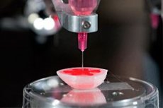
America has begun printing blood vessels using a 3D printer
Last reviewed: 02.07.2025

All iLive content is medically reviewed or fact checked to ensure as much factual accuracy as possible.
We have strict sourcing guidelines and only link to reputable media sites, academic research institutions and, whenever possible, medically peer reviewed studies. Note that the numbers in parentheses ([1], [2], etc.) are clickable links to these studies.
If you feel that any of our content is inaccurate, out-of-date, or otherwise questionable, please select it and press Ctrl + Enter.
Growing new human tissues in a laboratory environment is very difficult, because it is very painstaking and precise work. In addition to recreating natural structures, each tissue or organ must be artificially supplied with a vascular network, which is extremely difficult. If this is not done, the new tissue will not be able to receive nutrition and oxygen.
Specialists representing the University of California San Diego have developed a unique method of thin 3D printing of capillary and microvascular networks. The vessel walls are formed at a thickness of up to 600 microns.
The new technique is called "microscopic continuous optical biological printing." It will be used to recreate the vascular network for artificially grown organs or tissues with different structures.
The essence of the new method is as follows: cells of the required variety are immersed in a special hydrogel, after which, with the help of ultraviolet rays and temperature exposure, this mass is compacted, acquiring the required version of the three-dimensional structure.
Throughout the process, the cells remain alive and functionally capable: they then develop and fill the 3D framework.
During experiments on rodents, scientists transplanted artificially created vessels into experimental mice. At the same time, amazing results were demonstrated: new vessels completely took root after 14 days, and the wound surface healed much faster than usual.
The research was conducted under the supervision of nanoengineer Dr. Shaoshen Chen. According to him, this experiment allowed solving many problems of vascular biotechnology. Now it becomes clear how to recreate entire organs and individual tissues that would have a fully functional vascular network system. The issue of introducing vessels into individual parts of the body has also been clarified.
"The overwhelming majority of organs and tissues in the human body are permeated with blood vessels - this is necessary for the normal function and life of the organ. Vessels have always been considered the most vulnerable place in biotechnological and transplantation practice. Because of this, many scientific discoveries were not completed, and scientists were simply marking time. Now, the 3D printing of the vascular network that we have created will completely solve the previously arisen problem," Professor Chen commented on the discovery at a university press conference.
It is worth noting that Dr. Chen has been the head of the Nanobiomaterial, Biological Printing, and Tissue Biotechnology Lab at the University of California, San Diego for many years. He has been trying to recreate organs with full vascular filling for many years.
Today, scientists led by the professor continue their research. Now they have to improve the transport functionality of artificially created vessels. The specialists are also working on a new invention - the production of a vascular network from the patient's stem cells.
 [
[