New publications
AI-guided mammography reduces workload by 33% and increases detection of breast cancer
Last reviewed: 02.07.2025

All iLive content is medically reviewed or fact checked to ensure as much factual accuracy as possible.
We have strict sourcing guidelines and only link to reputable media sites, academic research institutions and, whenever possible, medically peer reviewed studies. Note that the numbers in parentheses ([1], [2], etc.) are clickable links to these studies.
If you feel that any of our content is inaccurate, out-of-date, or otherwise questionable, please select it and press Ctrl + Enter.
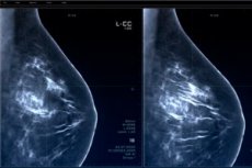
In a recent study published in the journal Radiology, researchers from Denmark and the Netherlands conducted a retrospective analysis of screening effectiveness and overall mammography screening burden before and after the introduction of artificial intelligence (AI) systems.
Regular mammography screening for breast cancer significantly reduces mortality from this disease. However, mass mammography screening increases the workload of radiologists who must analyze many mammograms, most of which do not contain suspicious lesions.
In addition, double screening, which is used to reduce false positives and improve detection, further increases the workload of radiologists. The shortage of specialized radiologists who can read mammograms exacerbates this situation.
Recent studies have extensively explored the use of AI to efficiently analyze radiology reports while maintaining high screening standards. A combined approach in which AI helps radiologists highlight mammograms with flagged lesions is thought to reduce the workload of radiologists while maintaining screening sensitivity.
The present study used preliminary performance measures from two cohorts of women undergoing mammography screening as part of the Danish national breast cancer screening program to compare the change in screening burden and performance after the introduction of AI tools.
The program invited women aged 50 to 69 to be screened every two years until age 79. Women with markers indicating an increased risk of breast cancer, such as the BRCA genes, were screened under different protocols.
The researchers used two cohorts of women: one screened before the AI system was introduced and one after. The analysis included only women under 70 years of age to exclude those in a high-risk subgroup.
All participants underwent standardized protocols using digital mammography with craniocaudal and mediolateral oblique views. All positive cases in this study were identified by screening for ductal carcinoma or invasive cancer, which were confirmed by needle biopsy. Data on pathology reports, lesion size, lymph node involvement, and diagnoses were also obtained from a national health registry.
The AI system used to analyze the mammograms was trained using deep learning models to detect, highlight, and score any suspicious calcifications or lumps on the mammogram. The AI then classified the screenings on a scale of 1 to 10, indicating the likelihood of breast cancer.
A team of mostly experienced radiologists reviewed mammograms for both cohorts. Before the AI system, each screening was reviewed by two radiologists, and a patient was recommended a clinical examination and needle biopsy only if both radiologists deemed the screening to require further evaluation.
After the AI system was implemented, mammograms with a score of 5 or less were reviewed by a senior radiologist, knowing that they would only receive one reading. Those that required further examination were discussed with a second radiologist.
The study found that the implementation of the AI system significantly reduced the workload of radiologists analyzing mammograms as part of mass breast cancer screening, while improving the effectiveness of screening.
The cohort screened before the AI system was implemented included over 60,000 women, while the cohort screened with AI included approximately 58,000 women. Screening with AI resulted in an increase in breast cancer diagnoses (0.70% before AI vs. 0.82% with AI) while reducing the number of false positives (2.39% vs. 1.63%).
AI-based screening had a higher positive predictive value and the percentage of invasive cancers was lower with AI-based methods. Although the percentage of node-negative cancers did not change, other performance measures showed that AI-based screening significantly improved outcomes. Reading load was also reduced by 33.5%.
In summary, the study assessed the effectiveness of an AI-based screening system in reducing radiologists' workload and improving screening rates in mammogram analysis as part of mass breast cancer screening in Denmark.
The results showed that the AI-based system significantly reduced the workload of radiologists while improving screening rates, as evidenced by a significant increase in breast cancer diagnoses and a significant reduction in false positives.