
New publications
Knocking out a single gene turned pancreatic duct cells into insulin-producing cells
Last updated: 09.09.2026

All iLive content is medically reviewed or fact checked to ensure as much factual accuracy as possible.
We have strict sourcing guidelines and only link to reputable media sites, academic research institutions and, whenever possible, medically peer reviewed studies. Note that the numbers in parentheses ([1], [2], etc.) are clickable links to these studies.
If you feel that any of our content is inaccurate, out-of-date, or otherwise questionable, please select it and press Ctrl + Enter.
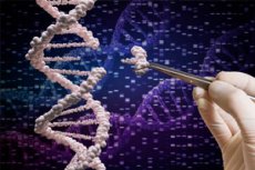
Scientists have discovered a genetic "brake" that, when turned off, allows human pancreatic duct cells to partially change their identity and transform into cells similar to insulin-producing β-cells. A study published September 2, 2026, in Science Translational Medicine identified the ALDH3B2 gene—aldehyde dehydrogenase 3 family member B2—as the regulator. [1]
To find this target, the researchers used a genome-wide CRISPR screen, sequentially knocking out genes in human pancreatic duct cells. The system encompassed over 19,000 genes. When ALDH3B2 was knocked out, the cells began to activate the insulin gene and other hallmarks of the β-cell program. In one functional measure, the frequency of β-like cells increased from less than 1% to approximately 8.5%.
The change was not merely molecular. The reprogrammed human cells secreted insulin in response to elevated glucose. After their transplantation into mice with experimental diabetes, glucose levels dropped to near normal and remained improved for about six weeks. When the transplant was removed, hyperglycemia returned, further indicating the functional role of the transplanted cells. [2]
However, a cure for diabetes in humans remains a long way off. The resulting cells were only β-like, not fully mature normal β cells: insulin secretion remained significantly weaker than in true human β cells, many cells continued to retain signs of ductal identity, and in vivo experiments have so far only been conducted on immunodeficient mice. Therefore, the work represents proof of principle, not a proven therapy. [3]
| Key fact | Result |
|---|---|
| The main target | ALDH3B2 |
| Type of study | preclinical, human cells + mouse model |
| Search method | whole-genome CRISPR screening |
| Gene tested | >19,000 |
| Source cells | human pancreatic duct cells |
| Result of ALDH3B2 shutdown | the emergence of a β-like phenotype |
| Estimated Conversion Rate | from <1% to ≈8.5% for one of the indicators |
| INS+ cells by single-cell RNA-seq | 18.1% versus 0.6% |
| KRT19 was preserved | approximately 93% of cells |
| Function | secretion of human insulin in response to glucose |
| Mouse model | streptozotocin-induced diabetes |
| Effect on glycemia | decrease to near normal for about 6 weeks |
| DOI | 10.1126/scitranslmed.ady2234 |
Why is β-cell restoration considered one of the main goals of diabetes treatment?
β-cells are located in the islets of Langerhans in the pancreas and perform a unique function: they constantly monitor blood glucose levels and secrete the exact amount of insulin required in response. This mechanism is significantly more complex than the simple presence of insulin in the body, as normal β-cells are capable of responding to changes in blood glucose levels in near real time.
In type 1 diabetes, β-cells are destroyed by the autoimmune system. In type 2 diabetes, the situation is more complex: insulin resistance, β-cell dysfunction, and a gradual decline in their functional mass develop simultaneously. Therefore, in both diseases, restoring a sufficient number of functioning β-cells is considered a potentially more radical strategy than simply compensating for the lack of insulin externally. [4]
Modern insulin therapy, continuous glucose monitoring systems, and automated insulin pumps can effectively control diabetes, but even the most advanced technologies cannot fully replicate the function of a healthy endocrine pancreas. Another approach—islet transplantation—can indeed restore endogenous insulin secretion, but is limited by donor shortages and the need for immunosuppression.
In recent years, β-cells derived from pluripotent stem cells have been actively developed. This approach has already reached early clinical trials. A new study proposes a fundamentally different approach: instead of cultivating new cells outside the body, attempting to transform the patient's existing ductal cells into new sources of insulin. Theoretically, such a strategy could utilize the patient's extremely accessible cellular reservoir.
| Approach | Advantage | The main problem |
|---|---|---|
| Insulin injections | effective glucose control | do not restore β-cells |
| Insulin pump | delivery automation | remains an external system |
| Donor islands | physiological secretion of insulin | donor shortage, immunosuppression |
| Stem cells | potentially unlimited source | immunity, production, safety |
| Ductal cell reprogramming | potentially the patient's own cells | so far only preclinical technology |
How scientists found ALDH3B2 among more than 19,000 genes
The first technical challenge was to create a system that would instantly indicate that a duct cell had begun to become β-like. The researchers used the human PANC-1 cell line and inserted a reporter construct linked to the insulin promoter. When the β-cell program was activated, the cell began to emit a green fluorescent signal.
Millions of these cells were then treated with a CRISPR-Cas9 library. Different cells received guide RNAs that silenced different genes. The idea was simple: if a gene normally prevents a ductal cell from transitioning to a β-cell state, its loss should lead to activation of the insulin reporter.
The screening covered virtually the entire human genome and identified several candidates. Among the eight most interesting genes, loss of ALDH3B2 resulted in the most significant activation of the insulin program, along with a reduction in some ductal identity features. The result was then confirmed with individual CRISPR constructs to rule out the random effect of a specific guide RNA. [5]
The researchers then moved from the immortal laboratory cell line to a much more important model—primary human pancreatic duct cells. Here, too, loss of ALDH3B2 function activated β-cell genes and led to the emergence of insulin-positive cells. This significantly enhances the translational value of the work: the phenomenon was not limited to a single tumor cell line. [6]
| Experimental stage | What did they do? |
|---|---|
| 1 | created cells with an insulin promoter reporter |
| 2 | applied the CRISPR library to the entire genome |
| 3 | looked for cells that activated a β-cell reporter |
| 4 | identified candidate genes |
| 5 | ALDH3B2 was switched off again using separate guides |
| 6 | confirmed the effect in primary human ductal cells |
| 7 | assessed RNA, proteins and insulin granules |
| 8 | tested the function after transplantation into mice |
The cells actually began to change their identity.
After loss of ALDH3B2, cells didn't simply begin synthesizing small amounts of insulin. A whole set of genes changed. Expression of INS, a key insulin gene, increased, as did transcriptional and functional markers of endocrine cells, including PDX1 and several genes involved in the production and processing of peptide hormones. Simultaneously, activity of some genes characteristic of the mature ductal phenotype decreased. [7]
Electron microscopy and immunohistochemistry also revealed intracellular granules containing insulin and C-peptide. The latter is particularly important as a sign that cells synthesize and process their own proinsulin, rather than simply absorbing pre-formed insulin from the external environment.
Using single-cell RNA sequencing, the researchers discovered an even more intriguing picture. 18.1% of cells with disrupted ALDH3B2 expressed INS, compared to only 0.6% of control cells. Among the INS-high cells, CHGA, IAPP, SCGN, SCG3, and SCG5—genes characteristic of endocrine and β-cells—were simultaneously elevated. [8]
However, approximately 93% of cells in both groups continued to express KRT19, a classic marker of ductal cells. This is a crucial detail: the transition was not complete. Many cells were in a hybrid state, combining features of ductal and endocrine identity. Therefore, the term "β-like cells" is significantly more accurate than the assertion that the researchers had already obtained fully functional normal β cells. [9]
Why do the numbers 8.5% and 18.1% appear?
| Indicator | Meaning | What does it mean? |
|---|---|---|
| Spontaneous conversion | <1% | rare occurrence of β-like phenotype without intervention |
| After ALDH3B2 shutdown | ≈8.5% | Conversion assessment using one of the functional reporting approaches |
| INS+ cells by scRNA-seq | 18.1% | cells in which insulin RNA was detected |
| INS+ in control | 0.6% | original background |
| KRT19+ | ≈93% | most cells retained some ductal identity |
These indicators do not contradict each other: they measure different characteristics and by different methods, and the presence of an INS transcript does not necessarily mean a fully mature functional β-cell.
Reprogramming even affected the cell's epigenetic memory.
One of the most interesting findings was changes in DNA methylation. In a normal ductal cell, the insulin gene is largely "closed." Chemical methylation marks on specific DNA regions help maintain this program and prevent the use of a gene that the ductal cell does not normally need.
After ALDH3B2 knockout, methylation levels at several well-studied sites within the human INS locus were significantly reduced. This effect was observed in both PANC-1 and primary human ductal cells. In other words, the intervention affected not only the short-term activity of RNA but also the epigenetic state of the insulin gene. [10]
This finding is potentially important for the stability of reprogramming. If a new cellular phenotype is maintained only by a short-term signaling cascade, the cell can quickly return to its original state after the stimulus is removed. INS demethylation indicates a more profound restructuring of the cellular program, although it alone does not prove the lifelong stability of the new phenotype.
The researchers also used single-cell analysis and differentiation trajectories. The model showed that cells likely do not transform directly from mature ductal cells to mature β-cells. They first partially lose their original identity and pass through an intermediate state resembling an immature pancreatic precursor, and then some begin to acquire endocrine and β-cell characteristics.
| Stage | Proposed cellular program |
|---|---|
| Original | mature ductal cell |
| Loss of ALDH3B2 | weakening of the stability of the ductal phenotype |
| Intermediate stage | pancreatic progenitor-like cell |
| Endocrine program | activation of hormonal genes |
| β-like state | INS, CHGA, IAPP and other markers |
| The final result | functional but incompletely mature β-like cell |
The most important test: do the new cells respond to glucose?
To potentially cure diabetes, simply forcing cells to produce insulin is not enough. Insulin must be secreted based on glucose concentration. Uncontrolled, continuous secretion would be potentially dangerous, as it could lead to severe hypoglycemia.
In laboratory tests, cells with knocked-out ALDH3B2 did indeed increase human insulin secretion when glucose levels rose. Insulin secretory granules were detected in them, and in transplantation experiments, human insulin appeared in the blood of mice after a glucose load. [11]
However, quantitatively, this response was significantly weaker than that of real human β-cells. This is one of the main limitations of the study. The cell can express INS, have granules, and even respond to glucose, but a fully functional mature β-cell utilizes an extremely complex system of glucose sensors, mitochondrial metabolism, ion channels, and exocytosis. The resulting cells so far only partially reproduce this system. [12]
Therefore, further improvement in efficiency will likely require additional factors. The authors themselves suggest that knocking out ALDH3B2 alone may not be sufficient to complete transdifferentiation. The next stage of research may involve identifying additional genetic or pharmacological interventions that promote the maturation of existing β-like cells. [13]
| A sign of a true β-cell | After ALDH3B2 shutdown |
|---|---|
| Insulin production | Yes |
| C-peptide | Yes |
| Secretory granules | Yes |
| Response to increased glucose | Yes |
| Expression of a number of β-cell genes | Yes |
| Complete loss of ductal markers | No |
| Secretion at the level of normal β-cells | No |
| Fully mature phenotype | Not yet |
Cell transplant lowers sugar levels in diabetic mice
To test the function in vivo, the researchers used immunodeficient mice in which diabetes was induced by streptozotocin, a substance that damages the body's own β-cells. This creates a model of severe insulin deficiency and allows them to test whether the transplant can compensate for the lost endocrine function. [14]
Human cells with knockout ALDH3B2 were transplanted into the kidney capsule—a classic experimental site for islet and β-cell transplantation. Control animals received cells without the desired genetic modification. Small groups of mice were used in the experiments described; in one series, approximately five animals were in each group. [15]
In animals receiving reprogrammed cells, glucose concentrations significantly decreased, approaching normal. The effect lasted for about six weeks, and human insulin could be detected in the bloodstream after a glucose load. This is one of the most powerful results of the study, as it demonstrates the functionality of cells beyond the petri dish. [16]
Further evidence was obtained upon transplant removal. After cell removal, around day 56, glucose levels rose again to levels typical of control diabetic animals. This links the improvement in glycemia specifically to the transplanted cells, rather than to the random restoration of the mouse's own pancreatic system. [17]
What the mouse model showed
| Indicator | ALDH3B2-deficient cells |
|---|---|
| Survived after transplantation | Yes |
| They produced human insulin | Yes |
| Reacted to glucose load | Yes |
| Reduced glycemia | Yes |
| Glucose level | was approaching normal |
| Duration of the observed effect | about 6 weeks |
| After removal of the graft | hyperglycemia returned |
Why ALDH3B2 Was Such an Unexpected Regulator
ALDH3B2 belongs to a large family of aldehyde dehydrogenases. These enzymes convert reactive aldehydes into the corresponding acids and are involved in protecting cells from toxic metabolic products. ALDH3B2 was previously linked to the metabolism of long-chain lipid aldehydes, but its role in maintaining pancreatic cell identity was largely unknown. [18]
It is particularly interesting that high ALDH activity in other tissues is often considered a characteristic of stem or progenitor cells. Different members of the family are used as markers of hematopoietic, neural, and potential pancreatic progenitors. However, different ALDHs have significantly different functions, so data on one enzyme cannot be automatically extrapolated to another. [19]
The authors have not yet definitively determined why the loss of ALDH3B2 increases ductal cell plasticity. A possible link is with lipid aldehyde metabolism, the cell's redox state, or metabolites that affect epigenetic enzymes. The observed demethylation of INS suggests that the link between ALDH3B2 metabolism and epigenetic state deserves further study. [20]
Importantly, the effect was not a universal property of all related aldehyde dehydrogenases. For example, disruption of the closely related ALDH3A1 did not reproduce the phenotype of ALDH3B2. This raises the possibility that the observed result is related to a specific function of ALDH3B2, rather than simply a general suppression of aldehyde metabolism. [21]
The most exciting prospect is that CRISPR may not be needed at all.
Although the discovery was made using CRISPR, ALDH3B2 itself is an enzyme. This is crucial: enzyme activity can often be inhibited by small molecules, meaning that theoretically, the same effect could be achieved with a drug without irreversible DNA editing.
The researchers tested this concept using DEAB, a broad inhibitor of some ALDHs. Exposure to DEAB also promoted the emergence of a β-like phenotype in human ductal cells. The authors view this as proof that this pharmacological approach is, in principle, feasible.
However, DEAB is not a specific inhibitor of ALDH3B2 and interacts with several members of the ALDH family. Therefore, it cannot be considered as a ready-to-use drug for β-cell restoration. Moreover, systemic inhibition of various ALDHs has the potential to impair the detoxification of toxic aldehydes in other organs. [22]
Therefore, the next important step is to create a selective inhibitor of ALDH3B2. If a molecule can be found that reaches pancreatic duct cells and temporarily inhibits this enzyme, researchers could theoretically test the idea of regenerating β-cells directly within the body without extracting, editing, and replanting the cells. However, such a drug does not yet exist. [23]
| Possible approach | Status |
|---|---|
| CRISPR knockout ALDH3B2 | works in cell experiments |
| Transplantation of modified cells | works in diabetic mice |
| Broad ALDH inhibitor DEAB | shows a similar effect in vitro |
| Specific ALDH3B2 inhibitor | still needs to be developed |
| β-cell regeneration tablet | for now it's a hypothetical prospect |
Could this strategy work for type 1 diabetes?
At first glance, the patient's own cells offer a significant advantage: they are genetically their own and therefore should not trigger the classic alloimmune rejection characteristic of donor cells. This potentially addresses one of the main challenges of islet transplantation.
But type 1 diabetes presents a more complex problem: autoimmunity is directed specifically against the β-cell phenotype. Even if a new β-like cell formed from the patient's own ductal epithelium, after the appearance of insulin and other β-cell antigens, it could theoretically become a new target of the same autoimmune system that destroyed the original β-cells.
The new study did not address this issue. The mice were immunodeficient specifically to prevent rejection of the human cells. Therefore, the results do not reflect how such cells would behave in a healthy human immune system, much less in active autoimmune diabetes. [24]
Therefore, in type 1 diabetes, restoration of β-cell mass will likely need to be combined with protection of new cells from autoimmune attack—immunomodulation, local protection, or other technologies. In type 2 diabetes, this barrier is potentially lower, but insulin resistance, metabolic stress, and a toxic environment remain, which can further impair the function of newly formed cells.
Why the results cannot yet be called a "cure for diabetes"
The first limitation is conversion efficiency. Even after ALDH3B2 intervention, not all ductal cells became β-like. Different methods yielded rates of approximately 8.5% or 18.1% INS-positive cells, with the vast majority continuing to express KRT19. This indicates partial and heterogeneous transdifferentiation.
The second limitation is maturity. The resulting cells produced significantly less insulin than normal human β-cells. Some cells simultaneously expressed multiple hormones, including INS, GCG, and PPY, reminiscent of immature endocrine states. There were also cells with features of the pancreatic progenitor phenotype. [25]
The third limitation is the mouse model. The animals were immunocompromised, the groups were small, the cells were transplanted under the renal capsule, and the observation period was weeks rather than years. It is unknown how long the new identity is maintained, whether the cells can proliferate uncontrollably, and what happens with long-term suppression of ALDH3B2 directly in the pancreas.
Finally, oncological risks must be excluded. Any intervention that allows a mature cell to lose its stable identity and revert to a more plastic progenitor-like state requires particularly careful evaluation. PANC-1 itself is a tumor-derived cell line, although key findings have also been confirmed in normal primary human ductal cells. Long-term evaluation of genomic stability, differentiation, and the potential formation of pathological cell populations will be necessary before clinical application. [26]
| What has already been shown | What else needs to be proven? |
|---|---|
| ALDH3B2 regulates duct cell plasticity | mechanism of action of ALDH3B2 |
| His loss includes INS | full β-cell maturity |
| Cells respond to glucose | secretion at the level of normal β-cells |
| Cells improve glycemia in mice | long-term effect |
| There is a pharmacological perspective | selective safe inhibitor |
| Human primary cells were used | efficiency inside the human pancreas |
| The patient's own cells are possible | protection against autoimmunity in type 1 diabetes |
| The effect lasts for weeks in mice. | safety for years |
How does this approach differ from stem cells?
Modern regenerative therapy for diabetes primarily attempts to generate new β-cells from embryonic or induced pluripotent stem cells. These cells are grown and differentiated outside the body, then transplanted into the patient. This approach offers a significant advantage—a potentially virtually unlimited supply of source material.
The ALDH3B2 strategy proposes an opposite philosophy: using the pancreas's own mature cells as a local source of new β-cells. The ductal system constitutes a significant portion of the exocrine pancreas, so even moderate conversion efficiency could potentially have biological significance if the process can be reliably triggered in vivo.
Theoretically, this approach could reduce dependence on cell factories, transplant procedures, and donor material. If a specific drug that targets only ALDH3B2 in the target cells is ever developed, regeneration could occur directly in the pancreas.
But technologically, this could prove even more difficult than transplantation. It's necessary to deliver the drug precisely to the target site, limit the extent of reprogramming, obtain the correct number of fully mature β-cells, and not disrupt the ductal structure. Therefore, the new strategy is currently opening a new avenue of research rather than competing with β-cell stem cell therapies already in clinical trials. [27]
What will scientists do next?
The first task is to understand the precise biochemical mechanism of ALDH3B2. While it is known that the enzyme is involved in aldehyde metabolism and its absence causes profound restructuring of cellular identity, it is unknown which specific substrate or product triggers this process. Finding such metabolites could explain the connection between metabolism and epigenetic switching in the cell. [28]
The second goal is to improve the quality of the resulting cells. Researchers need to achieve not just the appearance of INS, but a fully developed β-cell program: strong expression of PDX1, NKX6.1, MAFA, and other maturity factors, a proper glucose recognition system, and insulin secretion comparable to that of normal islets.
The third direction is the search for specific ALDH3B2 inhibitors. Because this is an enzyme, it is theoretically possible to use standard methods of medicinal chemistry and high-throughput screening of small molecules. The authors explicitly cite this pharmacological pathway as one potential way to translate the discovery into therapy. [29]
Only then will it be possible to move on to animal models that better mimic human diabetes, long-term experiments, and studies of the direct effects on endogenous pancreatic cells. The main question is whether it is possible to temporarily disable ALDH3B2 without removing cells from the body and obtain sufficient functional β-cells without tissue damage or unwanted reprogramming.
The main conclusion of the study
New work demonstrates that pancreatic cellular identity is more flexible than it might seem. Knocking down a single gene, ALDH3B2, was enough to trigger some human ductal cells to switch to a β-cell program, synthesize insulin, form secretory granules, and respond to glucose. [30]
Particularly compelling is the combination of several independent levels of evidence: whole-genome CRISPR screening, repeated genetic knockout of ALDH3B2, experiments with primary human cells, single-cell RNA sequencing, DNA methylation analysis, insulin secretion, and transplantation into diabetic mice.
At the same time, the study demonstrates how far we are from therapeutic potential. Most cells retained ductal features, functional secretion was weaker than that of normal β cells, and the safety and stability of the phenotype are still unknown. Therefore, this is more about "unlocking" the cell's hidden potential than a ready-made method for creating new, healthy β cell mass.
Nevertheless, the discovery of ALDH3B2 is of significant interest precisely because the target is an enzyme. If the effect can be reproduced with a specific, safe drug directly inside the pancreas, it could lead to a fundamentally new type of regenerative diabetes treatment—one that wouldn't rely on external insulin delivery or even cell transplantation, but rather on stimulating the body to create its own insulin-producing cells.
News source
Li J., Bode K., Lee Y.-C., Morrow N., Ma A., He M., Wei S., da Silva Pereira J., Stewart T., Lee-Papastavros A., Hollister-Lock J., Sullivan B.A., Pan H., Dreyfuss J.M., Bonner-Weir S., Yi P. Loss of function of ALDH3B2 transdifferentiates human pancreatic duct cells into β-like cells. Science Translational Medicine. 2026;18(865). Accessed September 2, 2026. DOI: 10.1126/scitranslmed.ady2234. PMID: 42685151.
The study is an original preclinical study, not a clinical trial. It utilized human cell lines, primary human pancreatic duct cells, whole-genome CRISPR screening, single-cell RNA sequencing, epigenetic studies, and transplantation of human cells into mice with experimental diabetes.