Medical expert of the article

New publications
Muscle ultrasound
Last reviewed: 20.11.2021

All iLive content is medically reviewed or fact checked to ensure as much factual accuracy as possible.
We have strict sourcing guidelines and only link to reputable media sites, academic research institutions and, whenever possible, medically peer reviewed studies. Note that the numbers in parentheses ([1], [2], etc.) are clickable links to these studies.
If you feel that any of our content is inaccurate, out-of-date, or otherwise questionable, please select it and press Ctrl + Enter.
About 30% of all sports injuries occur in the pathology of muscle tissue. Ultrasound examination is the leading one in diagnosing the pathology of muscle tissue, exceeding in resolution magnetic resonance imaging. In addition, the possibility of dynamic research in real time allows you to identify the invisible pathology in a static study.
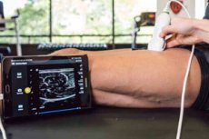
Ultrasound examination of muscle tissue (echography, ultrasound of muscles) is an informative diagnostic method, which is used to assess the state of soft tissues on almost any part of the human body. Muscle ultrasound is a simple and affordable method of examination, it allows you to assess the state of the tissue in real time.
Ultrasound procedure is completely harmless, if necessary, it can be repeated many times.
Indications for
Muscle tissue lesions are quite common in medical practice. The most common inflammatory processes on the background of diffuse connective tissue pathologies, congenital abnormalities, toxic muscle damage in cancer or hematological diseases, as well as myopathy, etc. It is not always advisable to use complex invasive studies, and not all patients have indications. Therefore, it is often the ultrasound of the muscles that becomes the selection procedure - a non-invasive diagnostic method that does not have a radiation effect and is relatively inexpensive (which is important).
Ultrasound of the muscles can be performed on almost any person: unlike tomographic procedures, ultrasound does not require complete long-term immobility of the patient, which is very important in relation to children and the elderly.
Ultrasound of the muscles helps to identify such pathological conditions as injuries, ruptures, hernias, hemorrhages, abscesses. You can also detect and various tumors: lipomas, cysts, sarcomas, liposarcomas, melanomas, glomus tumors, hemangiomas, neurofibromas, etc.
In addition, ultrasound of the muscles is used to clarify problem diagnoses, monitor the course of surgery, monitor the dynamics of treatment.
As a rule, this procedure is prescribed:
- with the appearance of pain in the muscles;
- with the forced restriction of motor activity;
- with and after injuries;
- with systemic pathologies;
- in acute inflammation (myositis);
- in the presence of edema or the appearance of palpable neoplasms of unknown origin.
 [1], [2], [3], [4], [5], [6], [7], [8], [9], [10], [11]
Necessary preparation
Special preparation for ultrasound of the muscles is not needed. If there are open skin lesions (wounds, scratches, cuts) at the site of the intended diagnostic manipulation, then it is necessary to wait for their healing.
Sometimes, with excessive hair growth in the examination area, it may be necessary to use a razor.
Other preparatory activities before the ultrasound of the muscles is not required. The patient can lead a normal life: there are no restrictions on food and fluid intake. It is desirable to come to the procedure in loose clothing, in order to be able to easily expose the part of the body being examined.
Detailed technique
Regardless of the location of the investigated area on the body, the technique of ultrasound of the muscles is always the same and consists of the following steps:
- The patient releases the required area on the body from the clothes.
- The patient fits on a couch, takes a comfortable position, relaxes.
- The doctor treats the skin at the site of examination with a special gel substance and applies an ultrasound probe.
- The doctor examines the affected tissue on the monitor screen: the resulting picture is the result of ultrasound reflection from the tissue surface.
At the end of the procedure, the gel substance should be removed with a napkin. Next, the patient is dressed and can go home. Additional care after the procedure is not required.
[15], [16], [17], [18], [19], [20], [21]
Contraindications to
The ultrasound procedure of the muscles has practically no contraindications: the diagnosis can be postponed if there are deep skin lesions, wounds, etc. On the body in the area of the intended examination. In general, this method is used in newborns, the elderly, and pregnant or lactating women.
Ultrasound of the muscles is well perceived by patients, because its implementation is not accompanied by any unpleasant sensations, and the study itself is short-term, safe and at the same time informative.
If necessary, ultrasound of the muscles can be repeated several times. For example, this is what happens when tracking the dynamics of tissue repair after surgical interventions and in some other pathologies.
What does muscle ultrasound show?
Healthy soft tissues for the most part have similar density and other characteristics. However, ultrasound of the muscles shows painful changes in the tissues more clearly and in detail, moreover in real time, and this is the main difference of this diagnostic method over other procedures.
Ultrasound of the muscles allows you to define even small pathological formations, which are fixed by the doctor on the monitor screen in the form of a change in the ECHO signal.
Most often, experts turn to scan the muscles of the following organs and body parts:
- Ultrasound of the leg muscles is performed to detect post-traumatic hematomas in the tissues of the thighs and ankle joint. The image of such seals has the form of localized foci with excessive blood filling. During the study, the doctor often asks the patient to move the limb in one direction or another: this allows us to consider the possible presence of a purulent process (on ultrasound, the density of the lesion changes at the moment of fluid displacement).
- Ultrasound of the thigh muscles is most often required after traumatic injuries, as well as if a tumor is suspected. If a patient has previously been diagnosed with a hernia of the hip joint, then an ultrasound method will help to assess the dynamics of treatment. In addition, the study is prescribed to clarify the nature of the manipulation before surgery, or to assess the condition of the tissue at its end.
- Ultrasound of the leg muscles is required for serious traumatic injuries of the ankle joint - and especially for suspected integrity of the muscles and / or tendons. More ultrasound helps detect tumor processes, cysts, and also allows you to monitor the quality of regeneration of damaged tissues.
- Ultrasound of the gastrocnemius muscle is usually recommended after injuries, as this method perfectly visualizes tissue breaks, small vessel lesions, hematomas. Any tumor processes (both benign and malignant) are also clearly visible.
- Ultrasound of the shoulder muscles is prescribed for degenerative changes in tissues, in the presence of an inflammatory process (arthritis, myositis), as well as for traumatic injury (stretching, rupture, contusion, hematoma, etc.). During the diagnosis, the doctor may ask the patient to raise his arm, take it to the side: changing the position of the limb allows you to more accurately assess the blood circulation in the area of tumor or inflammatory pathologies.
- Ultrasound of the abdominal muscles is mainly carried out to determine the tumor processes of various etiologies, to assess the state of blood circulation, to identify hemorrhages. Ultrasound can be applied in the postoperative period to monitor the dynamics of tissue healing.
- Ultrasound of the neck muscles is assigned to determine diseases of inflammatory etiology, to assess the area of muscle damage. Diagnosis is carried out when suspicious neoplasms in the form of balls, nodes, seals are palpated in the neck area. Additionally, during an ultrasound scan, the doctor may pay attention to the thyroid gland, carotid arteries, and the muscles that surround the trachea. When performing an ultrasound of the neck muscles, the doctor may ask the patient to turn his head, or tilt it slightly to the right or left.
- Ultrasound of the back muscles allows for a good view of the soft and cartilaginous tissue, as well as some bone tissues of the spine. Visualization of spinal structures, vasculature (you can determine the quality of blood circulation and blood supply) lend themselves perfectly. Ultrasound of the muscles is often used if the patient voices complaints of frequent headaches, limited movement in the neck or shoulder area, a feeling of crawling, numbness of the extremities, and dizziness.
- Ultrasound of the lower back muscles is important in the presence of aching pains extending to the lower extremities, with muscular numbness, with improper functioning of the organs located in the small pelvis. Ultrasound assists are especially often referred to when assessing the state of soft tissues after injuries and other damaging factors.
- Ultrasound of the pectoral muscle is prescribed for ruptures, osteophytes, myositis or hypoplasia / agenesis. Tears of the pectoral muscle are rarely met - with a direct blow to the chest, with a powerful eccentric muscle contraction. The ultrasound image of the pectoral muscle is a hypoechoic structure with echogenic septa perimisium inside. Often the study is practiced in the diagnosis of the condition of the muscles of the shoulder and / or thoracic spine.
- Ultrasound of nodal muscles is important mainly in children, but in some situations, the study is also carried out by adults - for example, with insufficient muscle blood supply, scarring and shortening as a result of muscular fiber tears. The second name for this type of diagnosis is ultrasound examination of the sternocleidomastoid muscle: such a oblique helix muscle runs through the cervical region from the mastoid process to the sternoclavicular joint. In adults, the injury of this muscle is relatively rare.
- Ultrasound of the piriformis muscle is performed with the same syndrome (referring to the piriformis syndrome): the structural changes of the sciatic nerve are studied (the line of the piriformis space and the distal direction to the bifurcation area). Diagnosis prescribed for pain in the gluteal zones, with the spread of pain on the lower limbs or perineum, with numbness of the plantar region.
- Ultrasound of the arm muscles is used for a detailed examination of suspicious tumors - not only in the area of muscles, but also in the joints, vessels. Often, patients with complaints of regular aching pain in the limb, limitation of mobility, which is not associated with problems in the joints, appeal for such a diagnosis. After an injury, an ultrasound scan will indicate the nature and extent of damage to the arm muscles.
- Ultrasound of the trapezius muscle is prescribed in case of its overstrain, stretching due to high-intensity workouts, as well as in case of bruise, miglosis, idiopathic pain. The study allows to establish the correct diagnosis, if the essence of the disease can not be established by the method of ordinary palpation.
- Ultrasound of the masticatory muscles is most often prescribed to assess the effects of their traumatic damage. Immediately after the injury, the study will help determine the size of the hematoma. In addition, such a diagnosis is carried out in the presence of purulent or other neoplasms and nodes in the facial area.
- Ultrasound of the kivatal muscles in children is carried out with congenital insufficient development of the sternocleidomastoid muscle, after traumatizing it during labor, and also with birth injury to the cervical vertebral part. Ultrasound of the muscles determines the inflammatory changes in the tissues, is used to diagnose tumors. Especially often the procedure allows to identify torticollis, as well as determine the functionality of arterial vessels that supply blood to the brain.
- Ultrasound of the eye muscles helps to investigate the quality of eyeball movements, assess the structure of the eye muscles and optic nerve, identify tumors, strictures, effusion, etc. In addition, ultrasound is able to determine the pathological changes in the eye blood circulation at the initial stage of development. This type of diagnosis is not carried out with the wound of the eyelids and the peri-orbital zone, with open traumatic eye damage, with retrobulbar bleeding.
Reviews
Negative reviews about this diagnostic method, like ultrasound of the muscles, are practically absent. It is an inexpensive, safe and highly accurate way to detect various neoplasms and inflammatory changes. The procedure allows to assess the probability of post-traumatic consequences, to detect foreign bodies in muscle tissues.
Muscle pathologies on ultrasound are manifested by a change in the structure of tissues, an increased acoustic density, a pronounced change in blood circulation in muscle tissue during exercise. Tissues are reliably visualized, the characteristic features of the structure of the muscles are determined depending on the age of the patient.
Muscle ultrasound is a simple and affordable diagnosis that is highly informative. Unlike many other studies, this procedure can be repeated many times without any harm to health. Especially often this method is used in traumatology and emergency medicine, as well as to identify tumor processes.