Medical expert of the article

New publications
Temporal bone
Last reviewed: 19.11.2021

All iLive content is medically reviewed or fact checked to ensure as much factual accuracy as possible.
We have strict sourcing guidelines and only link to reputable media sites, academic research institutions and, whenever possible, medically peer reviewed studies. Note that the numbers in parentheses ([1], [2], etc.) are clickable links to these studies.
If you feel that any of our content is inaccurate, out-of-date, or otherwise questionable, please select it and press Ctrl + Enter.
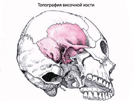
The temporal bone (os temporale) is paired, it forms part of the base and lateral wall of the skull between the sphenoid bone in front and the occipital bone behind. It accommodates the organs of hearing and balance. In the temporal bone, a pyramid, a drum and a scaly part are distinguished.
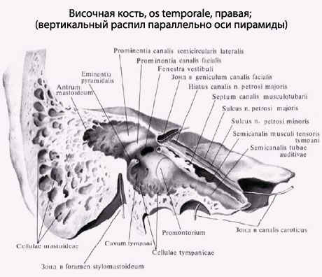
The pyramid, or rocky part (pars petrosa), has a triangular shape, located obliquely in the horizontal plane. The tip of the pyramid is directed forward and medially, and the base is back and lateral. At the top of the pyramid is the inner opening of the carotid canal (canalis caroticus). Nearby and lateral is the musculoskeletal canal (canalis musculotubarius), which is divided into two half-channels by a septum: semicanalis tubae auditivae and semicanalis musculi tensoris tympani muscle.
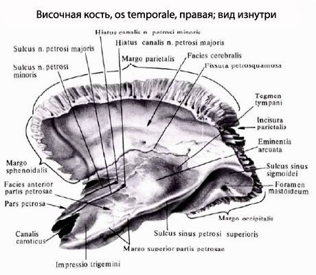
The pyramid is distinguished by three surfaces: the front, back and bottom. The front surface of the pyramid faces up and forward. Near the top on this surface there is a slight trigeminal impression (impressio trigemini). Two openings are noticeable laterally than this depression. The larger of these is called the cleft (orifice) of the canal of the large stony nerve (hiatus canalis nervi petrosi majoris), from which a narrow furrow with the same name extends forward and medially. A cleft of a small stony nerve (hiatus canalis nervi petrosi minoris), leading to the furrow of this nerve, lies anterior and lateral. On the front surface of the pyramid there is a flattened section - the roof of the drum cavity (tegmen thympani), which is its upper wall. Along the upper edge of the pyramid is a groove of the upper stony sinus (sulcus sinus petrosi superioris).
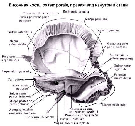
The posterior surface of the pyramid faces posteriorly and medially. On the middle of this surface is the inner auditory opening (porus acusticus internus). It leads to the internal auditory meatus (medtus acusticus internus). Lateral and somewhat above this opening is the fossa subarcuata, below, and laterally the slightly noticeable external aperture (hole) of the aqueduct of the vestibule (apertura externa aqueductus vestibuli). Along the posterior edge of the pyramid is a groove of the lower stony sinus (sulcus sinus petrosi inferioris). At the lateral end of this furrow, next to the jugular fossa, there is a depression at the bottom of which opens the outer aperture of the tubule of the cochlea (apertura externa canaliculi cochleae).
The lower surface of the pyramid has a complex relief. Near the base of the pyramid is a deep jugular fossa (fossa jugularis). Ahead of her is a rounded outer opening of the canal canal, inside which, in its wall, there are 2-3 holes of the sleepy-tympanic tubule, connecting the sleeping canal with the drum cavity. On the scallop between the jugular fossa and the external opening of the carotid canal is a small lobe (fossula petrosa). Lateral to the jugular fossa, a thin and long subulate process (processus styloideus) is directed downward. Behind the appendage there is a stylomastoid aperture (foramen stylomastoideum), and behind this opening is directed down a wide, easily mastoidal process (mastoidus) that is easily palpable through the skin.
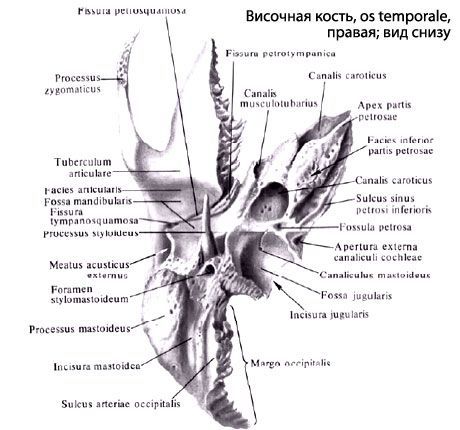
In the thick of the mastoid process there are air-filled cells. The most compartmental cell - the mastoid cave (Antrum mastoideum) communicates with the drum cavity. Medially mastoid process is limited by a deep mastoid fillet (incisure mastoidea). The median of this notch is the furrow of the occipital artery (sulcus arteriae occipitalis). At the base of the mastoid process there is sometimes a mastoid opening (foramen mastoideum).
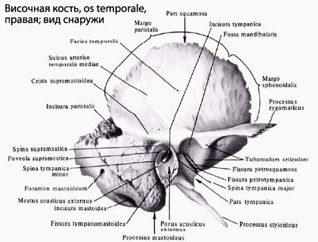
The drum part (pars tympanica) is formed by a curved narrow bone plate, which in front, below and behind restricts the external auditory opening (porus acusticus externus) leading to the external auditory meatus (meatus acusticus externus). Between the drum part and the mastoid process is a narrow drum-mastoid fissure (fissure tympanomastoidea). In front of the external auditory aperture there is a tympanosquamosa (fissure tympanosquamosa). In this gap from inside comes a narrow bone plate - the edge of the roof of the drum cavity. As a result, the drum-scaly slit is divided into an anteriorly stony-scaly slit (fissura petrosquamosa) and a stony-drum slit (fissura petrotympanica, an eyelet slit) through which a branch of the facial nerve emerges from the tympanic cavity - a drum string.
The scaly part (pars squamosa) is a convex outwardly shaped plate having a beveled free upper edge for connection to the parietal bone and a large wing of the sphenoid bone. The external temporal surface of the scales is smooth. On the inner cerebral surface of the scales there are cerebral elevations, finger-like impressions and arterial furrows. From the scales, above and anterior to the external auditory canal, the zygomatic process (processus zygomaticus) begins. Connecting with the temporal process of the malar bone, it forms a zygomatic arch. Behind the zygomatic process, at its base, is the mandibular fossa (fossa mandibularis) for articulation with the condylar process of the lower jaw to form the temporomandibular joint.
Channels of the temporal bone. Several channels of the temporal bone pass through the pyramid for cranial nerves and blood vessels.
The canalis canalis cardticus) begins on the lower surface of the pyramid with an external carotid opening, goes upward, bends almost at right angles, then is guided medially and forward. The canal ends with an internal carotid opening at the apex of the temporal bone pyramid. Through this channel, the internal carotid artery and the nerves of the carotid plexus pass into the cavity of the skull.
Sleepy-drum canals (canaliculi caroticotympanic!), Number 2-3, depart from the sleeping canal and are sent to the tympanum. In these tubules are located the same name arteries and nerves.
The musculoskeletal canal (canalis musculotubarius) begins at the apex of the pyramid of the temporal bone, goes back and laterally and opens into the tympanum. The horizontal partition divides it into two parts. Above is the seminal canal of the muscle that strains the eardrum (semicanalis musculi tensoris tympani) containing the same muscle. Below is the seminal duct of the auditory tube (semicanalis tubae auditivae).
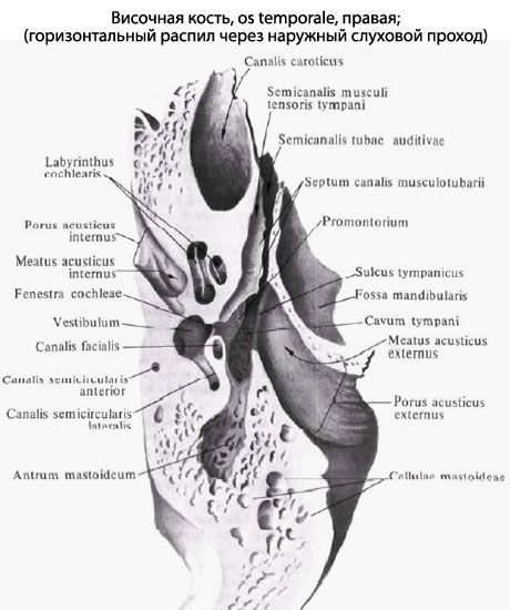
The canalis canalis facialis begins in the internal auditory canal. It goes first transversely with respect to the long axis of the pyramid to the level of the cleft of the canal of the large stony nerve. Having reached the cleft, the channel forms a knee, then it is guided at a right angle back and laterally. Passing along the medial wall of the tympanic cavity, the canal turns vertically downward and ends with a stylus-like aperture. In this canal passes the facial nerve.
The canaliculus of the drum string (canaliculus chordae tympani) comes from the wall of the facial canal in its final section and opens into the tympanum. In this channel there is a nerve - a drum string.
The drum canaliculus (canaliculus tympanicus) begins at the bottom of the stony dimple, it goes up, perforates the wall of the tympanic cavity. Further, the tubule passes along its medial wall and ends in the area of the cleft of the channel of the small stony nerve. In this tubule passes the drum nerve.
The canalic canaliculus (canaliculus mastoideus) begins in the jugular fossa and terminates in the tympoid-mastoid fissure. In this tubule passes the ear branch of the vagus nerve.