Medical expert of the article

New publications
Faringomicosis
Last reviewed: 23.04.2024

All iLive content is medically reviewed or fact checked to ensure as much factual accuracy as possible.
We have strict sourcing guidelines and only link to reputable media sites, academic research institutions and, whenever possible, medically peer reviewed studies. Note that the numbers in parentheses ([1], [2], etc.) are clickable links to these studies.
If you feel that any of our content is inaccurate, out-of-date, or otherwise questionable, please select it and press Ctrl + Enter.
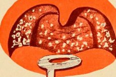
Pharyngomycosis (tonsillomycosis, fungal infection of the oral cavity, fungal pharyngitis, fungal tonsillitis, fungal infection of the pharynx, thrush) - pharyngitis (tonsillitis) caused by fungi. Pharyngitis is an inflammation of the mucous membrane of the oropharynx. Tonsillitis is an inflammation of one or more lymphoid formations of the pharyngeal coli, most often of the tonsils. In most cases, the disease is caused by yeast-like, less mold fungi.
Epidemiology
The frequency of pharyngomycosis over the past 10 years has increased dramatically and is 30-45% in the structure of infectious lesions of the pharynx and tonsils. The increase in the number of patients with this pathology is due to a significant increase in the number of risk factors for their development, among which the leading positions are taken by iatrogenic immunodeficiency states resulting from massive antibacterial therapy, prolonged use of glucocorticoid and immunosuppressive drugs for cancer, blood diseases, HIV infection, endocrinopathy, H Such situations have all the prerequisites for the development of pharyngomycosis, because the causative agents of the disease are opportunistic fungi. S, saprophytic on the mucous membrane of the oropharynx and in the environment.
The problem of pharyngomycosis acquires an important social significance not only because of the increasing distribution, but also because the fungal infection of the oropharynx is more severe than other inflammatory processes of this localization. A fungal infection of the oropharynx may be the primary focus of disseminated visceral mycosis or the cause of fungal sepsis.
In childhood, the incidence of pharyngomycosis is high. Especially common candidiasis of the oral mucosa in newborns (thrush). The occurrence of candidiasis is associated with the incompleteness of the formation of immune protection in newborns from the effects of mycotic infection. Pharyngomycosis often affects older children. In many of them, the onset of the disease is associated with fungal infection at an early age and incomplete elimination of the pathogen from the source of infection.
In the adult population, mycosis of the pharynx is diagnosed with the same frequency between the ages of 16 and 70 years, and in some cases even at an older age.
Causes of the pharyngomycosis
The main causative agents of pharyngomycosis are considered to be various types of yeast-like fungi of the genus Candida (in 93% of cases): C. Albicans, C. Tropicalis, C.krusei, C. Glabrata, C. Parapsillosis, S. Stellatoidea, C. Intermedia, S. Brumpti, C sake et al. C. Albicans (in 50% of cases) is considered the main causative agent, C. Stellatoidea ranks second in frequency of occurrence. This species is similar in morphological and biochemical properties to C. Albicans, and many authors identify them.
In 5% of cases, fungal lesions of the oropharynx are caused by mold fungi of the genera Geotrichum, Aspergillus, Penicillium, etc.
Pathogenesis
The main causative agents of pharyngomycosis are considered to be various types of yeast-like fungi of the genus Candida (in 93% of cases): C. Albicans, C. Tropicalis, C.krusei, C. Glabrata, C. Parapsillosis, S. Stellatoidea, C. Intermedia, S. Brumpti, C sake et al. C. Albicans (in 50% of cases) is considered the main causative agent, C. Stellatoidea ranks second in frequency of occurrence. This species is similar in morphological and biochemical properties to C. Albicans, and many authors identify them.
In 5% of cases, fungal lesions of the oropharynx are caused by mold fungi of the genera Geotrichum, Aspergillus, Penicillium, etc.
Symptoms of the pharyngomycosis
With pharyngomycosis, patients complain of discomfort in the throat, burning sensation, dryness, soreness, tickling, which are more pronounced than in bacterial lesions of the pharynx. The pain is moderate in intensity, when swallowing and ingestion of irritating food intensifies. Patients noted the irradiation of pain in the submandibular region, on the front surface of the neck and in the ear. The specific signs of pharyngomycosis are the detection of plaque, edema of the mucous membrane and pronounced intoxication phenomena. Also, pharyngomycosis is characterized by frequent exacerbations (2-10 times a year) and the development of the disease at any age.
The clinical course of pharyngomycosis can be acute and chronic. The process is localized mainly on the tonsils, the palatine arches, the back of the throat. Patients have a feeling of scratching, burning and discomfort in the pharynx, malaise, headache, low-grade fever. With pharyngomycosis caused by yeast-like fungi, whitish patches of various sizes are found in the pharynx, which are easily removed, exposing the hyperemic mucous membrane areas, less often bleeding ulcerations. Pharyngomycosis caused by mold fungi, characterized by the fact that the raids are yellowish, difficult to remove, which may cause suspicion of the presence of diphtheria of the throat. It is possible to spread fungi to the larynx, esophagus, the formation of paratonsillar abscesses.
What's bothering you?
Forms
In accordance with the localization of mycotic lesions, emit:
- cheilite;
- glossit;
- stomatitis;
- gingivitis;
- tonsillitis
- pharyngitis.
According to the clinical course, the following forms of pharyngomycosis are distinguished:
- acute:
- chronic.
In many cases, the acute process becomes chronic due to improper diagnosis and irrational treatment.
Clinical and morphological variants of pharyngomycosis:
- pseudomembranous. It is characterized by white patches of a cheesy appearance, which are stripped off with a bright red base, sometimes with a bleeding surface:
- erythematous (catarrhal). Characterized by erythema with a smooth "lacquered" surface, while patients note pain, burning, dryness in the mouth;
- hyperplastic. In the oral cavity, white spots and plaques are found that are difficult to separate from the underlying epithelium;
- erosive and ulcerative.
Diagnostics of the pharyngomycosis
During the survey, the following data are necessarily taken into account: time of onset of the disease, features of the course. It is necessary to find out whether the patient had previously had paratonsillitis and paratonsillar abscesses, frequency, duration, and nature of exacerbations of tonsillitis. Consider the previous treatment (local or general), its effectiveness. It is imperative to find out whether the patient was treated with antibiotics, glucocorticoids, cytostatics (duration and intensity of treatment), especially industrial and household conditions, previous diseases, allergic history. It should be borne in mind that in patients with pharyngomycosis, frequent exacerbations, absence or little effect from standard treatment methods are noted.
Physical examination
On examination, the following morphological changes are detected: infiltration of the mucous membrane, dilation and injection of blood vessels, desquamation of the epithelium. A characteristic clinical sign of chronic pharyngitis of fungal etiology is considered uneven hyperemia and infiltration of the mucous membrane of the posterior pharyngeal wall. Against the background of subatrophy, an increase in lateral cushions is noted. Often, on the background of the described pathological changes, whitish, cheesy, easily removed scurfs are revealed, under which they reveal erosion sites of the mucous membrane. In the case of ulcerous-necrotic form of fungal tonsillitis, the raids extend beyond the palatine tonsils to the palatine arches and the soft, and sometimes hard palate. Detection of plaque and unilateral damage is considered pathognomonic diagnostic signs of pharyngomycosis.
In chronic tonsillitis, examination is carried out outside the period of exacerbation. It is necessary to pay attention to the color of the mucous membrane of the oropharynx, tonsils, the nature of the raids (their color, prevalence), the size of the tonsils, the degree of swelling, consistency (dense or loose), cohesion with the arms, the presence of purulent contents in the gaps. Be sure to inspect the lingual tonsil (pay attention to its color, size, presence of raids), lymph nodes.
 [17]
[17]
Laboratory research
Fungal lesion of the pharynx may be suspected on the basis of endoscopic data, but mycological laboratory research methods are crucial for making a correct diagnosis. At the same time once received negative results do not indicate the absence of a fungal disease, therefore in this situation it is necessary to conduct repeated studies of the pathological discharge. At the same time, a single growth of fungi in crops does not always indicate a fungal infection.
When mycological examination is carried out microscopy, and then the seeding of the pathological discharge on nutrient medium. For accurate diagnosis is important the correct collection of pathological material for research. Targets from the surface of the tonsils are usually easily removed. Large, dense raids are removed on a glass slide with ear forceps and, without spreading, covered with another glass slide. Lean scurfs are removed with a Volkmam spoon, carefully so as not to injure the tissue.
When candidiasis of the tonsils is important microscopic examination of both native and colored drug. When coloring according to Romanovsky-Giemsa, spores of yeast-like fungi of the genus Candida are revealed. Cells of the fungus are round or elongated, the process of budding is clearly visible, as well as the threads of the pseudomycelium. Mycelium of yeast-like fungi of the genus Candida consists of bundles of elongated cells, connected in chains, which resemble a true mycelium. A true mycelium is a long tube divided by transverse partitions with a single shell. Pseudomycelium does not have a common shell. The morphological features of the fungus of the genus Candida are considered to be one of the reliable features that distinguish it from other fungi.
In the initial stage of the disease, microscopy of plaque reveals accumulations of fungal blastospores, and the threads of the pseudo-mycelium are isolated or absent. In the midst of the disease, clusters of budding fungus cells and numerous threads of pseudomycelium are visible in the smear. Thus, an accurate diagnosis can be established already according to microscopic examination.
Cultural studies are considered one of the important methods for the diagnosis of candidiasis. Using these methods, not only confirm the diagnosis of a fungal disease, but also determine the type of pathogen, judge the effectiveness of treatment.
When sowing on elective environments in patients with pharyngomycosis, yeast-like fungi of the genus Candida are most often isolated. When sowing on Saburo medium, uniform growth of yeast-like fungi of the genus Candida is observed at each planting point (to exclude errors, sowing is carried out in 2-4 tubes).
In chronic tonsillitis, when there is no plaque, seeding is done in the following manner. Material for seeding is taken from both tonsils and the posterior pharyngeal wall with a sterile cotton swab. The tampons are placed in sterile tubes with liquid Saburo fluid, and then in a thermostat for 24 hours at an ambient temperature of 27-28 C. After that, the material is subcultured onto the Saburo solid medium simultaneously in 3 tubes. After reseeding, the tubes are again placed in a thermostat for 8-10 days. Already on the 4-5th day, the fungi of the genus Candida give a characteristic growth of the colonies round, white or whitish-gray, their surface is convex, smooth and shiny, the texture is curdled.
If the fungi are found in the microscopic examination of the tonsils, they can also be distinguished when planted in pure culture. As a rule, continuous growth is noted (30-45 thousand colonies in 1 ml).
In addition, clinical blood tests (including HIV infection, markers of hepatitis, syphilis), urine must be done, urine must be determined, blood glucose levels, and immunogram parameters must be determined.
Thus, the diagnosis of fungal infections of the pharynx is based on:
- clinical data;
- detection of fungi by microscopy of smears from the mucous membrane;
- positive results when sowing on elective nutrient media.
Screening
Screening method for the detection of pharyngomycosis is a microscopy of a native and colored smear preparation from the mucous membrane of the pharyngeal cavity and the surface of the tonsils.
What do need to examine?
How to examine?
What tests are needed?
Differential diagnosis
Differential diagnostics should be performed with acute bacterial pharyngitis and tonsillitis, scarlet fever, diphtheria, tuberculosis, syphilis, anginal form of infectious mononucleosis, angina Simanovsky-Plut-Vincent, malignant neoplasms.
Indications for consulting other specialists
Consultation of an immunologist is needed to identify and correct immunodeficiency states; endocrinologist - to identify endocrine pathology, correction of endocrinopathy; oncologist - to exclude tumors of the oral cavity and pharynx; infectious diseases - to exclude diphtheria and mononucleosis.
Who to contact?
Treatment of the pharyngomycosis
The treatment is aimed at eliminating the fungus-pathogens and correction of the immunodeficiency state.
Indications for hospitalization
Complicated forms of pharyngomycosis.
Drug treatment of pharyngomycosis
General principles of pharmacotherapy of fungal infections of the oropharynx:
- The use of systemic antifungal drugs must be combined with a local effect on the site of infection;
- Antifungal drug therapy should be based on the results of a laboratory study of the sensitivity of the fungus to the drug used.
The treatment of pharyngomycosis consists in prescribing the following drugs: nystatin tablets, which are chewed and enveloped in the received mass with the surface of the pharynx with movements of the tongue and swallowing movements. With inefficiency - levorin, dekamin. The lesions are smeared with 1% solution of gentian violet, 10% solution of sodium tetraborate in glycerol, Lugol solution.
With the ineffectiveness of treatment with standard doses of fluconazole, itraconazole is prescribed 100 mg per day or ketoconazole 200 mg per day for a month. Itraconazole acts not only on yeast-like fungi of the genus Candida, but also on mold fungi.
For pharyngomycosis resistant to other antimycotics, amphotericin B is administered intravenously at 0.3 mg / kg per day for 3–7 days. Treatment of pharyngomycosis with amphotericin B and ketoconazole is carried out under the control of biochemical parameters of liver and kidney functions, since these drugs, especially amphotericin B, have a pronounced nephro- and hepatotoxic effect.
For systemic treatment of pharyngomycosis, drugs of the following antimycotic groups are used:
- polyenes: amphotericin B, nystatin, levorin, natamycin:
- azole: fluconazole, itraconazole, ketoconazole;
- allylamine: terbinafine.
Fluconazole is the most effective for pharyngomycosis, which is prescribed 1 time per day in a dose of 50 or 100 mg, in severe cases - 200 mg. The course of treatment is 7-14 days.
Alternative treatment regimens for pharyngomycosis, also lasting 7-14 days, consider the following;
- Levorin suspension (20 000 U / ml) 10-20 ml 3-4 times a day; "Natamycin suspension (2.5%), 1 ml 4-6 times a day;
- suspension of nystatin (100 000 U / ml) 5-10 ml 4 times a day.
With the ineffectiveness of treatment with standard doses of fluconazole, itraconazole is prescribed 100 mg per day or ketoconazole 200 mg per day for a month. Itraconazole acts not only on yeast-like fungi of the genus Candida, but also on mold fungi.
For pharyngomycosis resistant to other antimycotics, amphotericin B is administered intravenously at 0.3 mg / kg per day for 3–7 days. Treatment with amphotericin B and ketoconazole is carried out under the control of biochemical parameters of the functions of the liver and kidneys, as these drugs, especially amphotericin B, have a pronounced nephrotoxic and hepatotoxic effect.
In case of mold mycoses, itraconazole and terbinafine are considered the most effective. The course of treatment with itraconazole is 14 days, 100 mg 1 time per day, terbinafine - 8-16 days, 250 mg 1 time per day.
With topical treatment, antiseptics and antimycotics (Miramistin, hydroxyquinoline, clotrimazole, borax in glycerin, natamycin suspension) are used for lubrication, rinsing, irrigation, washing the lacunae of the tonsils.
Antimycotics are used against the background of elimination of risk factors, for example, neutropenia, careful treatment of dentures, etc.
Further management
In case of exacerbation of pharyngomycosis, the azoles are prescribed by mouth or topically for 7-14 days, taking into account the drug sensitivity of the pathogen. Need to eliminate risk factors. After achieving remission, anti-relapse treatment with systemic antimycotics or antifungal drugs for topical administration is performed.
More information of the treatment
Prevention
The main measures for the prevention of pharyngomycosis should be directed to the elimination of factors contributing to the activation of fungal flora, namely, the abolition of antibiotics, glucocorticoids, correction of the glycemic profiles fortifying therapy.
Forecast
With timely treatment and adequate antifungal therapy, the prognosis is favorable. Approximate terms of disability during exacerbation of pharyngomycosis is 7-14 days.