Medical expert of the article

New publications
Bariatric surgery
Last reviewed: 17.10.2021

All iLive content is medically reviewed or fact checked to ensure as much factual accuracy as possible.
We have strict sourcing guidelines and only link to reputable media sites, academic research institutions and, whenever possible, medically peer reviewed studies. Note that the numbers in parentheses ([1], [2], etc.) are clickable links to these studies.
If you feel that any of our content is inaccurate, out-of-date, or otherwise questionable, please select it and press Ctrl + Enter.
Bariatric surgery is a branch of medicine that studies the causes, treatment and prevention of obesity.
The term "bariatria" comes from the Greek words "baros" - weight and "yatrik" - treatment. This term has been applied since 1965. Bariatria includes both medication and surgical treatment.
Bariatric surgery
The "golden standard" in bariatric surgery is three types of operations:
- the introduction of an intragastric balloon (which, strictly speaking, is not an operation - is an outpatient endoscopic procedure)
- gastric banding operation
- gastric bypass surgery
According to modern requirements, all bariatric operations should be performed exclusively laparoscopically - i.e. Without wide surgical incisions. This technology can significantly ease the postoperative period and reduce the risk of postoperative complications.
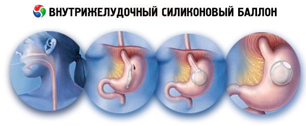
Intragastric silicone balloon
The installation of the intragastric balloon is referred to the group of gastrestrictive interventions. These cylinders are designed to reduce body weight, the mechanism of their action is based on a decrease in the volume of the stomach cavity when it is introduced into the latter, which leads to a faster formation of a feeling of saturation due to a partial (reduced) filling of the stomach with food.
The balloon is filled with saline solution, due to which it takes a spherical shape. The balloon freely moves into the cavity of the stomach. Adjust the filling of the balloon is possible within 400 - 800 cm 3. Self-closing valve allows you to isolate the balloon from external catheters. The balloon is placed inside the catheter unit, designed to insert the balloon itself. The catheter block consists of a silicone tube with a diameter of 6.5 mm, one end of which is connected to a shell containing a blown canister. The other end of the tube fits the special Luer-Lock cone connected to the balloon filling system. The catheter tube has risks for controlling the length of the injected part of the catheter. To increase the stiffness, a conductor is placed inside the hollow tube. The filling system in turn consists of a T-shaped tip. The filling tube and the filling valve.
According to the literature, different authors give different indications for the installation of an intragastric balloon for correction of obesity and overweight. We consider it advisable to treat with this technique always when there are no contraindications.
Contraindications to the use of an intragastric balloon
- diseases of the gastrointestinal tract;
- severe cardiopulmonary diseases;
- alcoholism, drug addiction;
- age is less than 18 years;
- presence of chronic foci of infection;
- unwillingness or inability to comply with the patient's diet;
- emotional instability or any psychological qualities of the patient, which, according to the surgeon, make the application of this method of treatment undesirable.
With BMI (body mass index) less than 35, the intragastric balloon is used as an independent treatment method, with a BMI over 45 (overcrossing), the intragastric balloon is used as preparation for the subsequent operation.
Intragastric silicone balloon is intended for temporary use in the treatment of patients suffering from overweight and obesity. The maximum period during which the system can be in the stomach is 6 months. After this period, the system must be removed. When the balloon is kept in the stomach for a longer time, the gastric juice acting on the cylinder wall destroys the latter, the filler leakage, the balloon decreases in size, as a result of which the balloon can migrate into the intestine with the onset of acute intestinal obstruction.
 [5], [6], [7], [8], [9], [10], [11], [12]
The method of mounting the cylinder
After a standard premedication, the patient in the endoscopic cabinet is placed on the left side. Intravenous sedative (Relanium) is administered. A probe is inserted into the esophagus with a balloon attached to it. Then a fibrogastroscope is inserted into the stomach and the presence of a balloon in its cavity is visually confirmed, the conductor is removed from the probe and the balloon is filled with a sterile saline solution of sodium chloride.
The liquid should be injected slowly and evenly to avoid rupture of the balloon. On average, the volume to be filled should be 600 ml, while the gastric cavity should remain free. After filling the balloon fibrogastroscope is carried to the esophagus to the level of cardiac pulp, the balloon is pulled to the cardia and the probe is removed from the nipple valve. In this case, with the help of a fibrogastroscope, the cylinder's traction is created in the opposite direction, which facilitates the removal of the conductor.
After removing the probe itself, the balloon is inspected for leaks. The balloon can be installed on an outpatient basis in an endoscopic room, without hospitalization in the hospital.
The method of removing the cylinder
The cylinder is removed under the condition of complete evacuation of the liquid from it. For this, a special tool is used, consisting of a needle with a diameter of 1.2 mm, reinforced on a long rigid conductor - a string. This perforator is carried out along a fibrogastroscope channel into the stomach at an angle of 90 degrees to the balloon. The balloon moves to the antrum of the stomach and becomes more accessible for manipulation. Then the cylinder wall is perforated. The conductor with the needle is removed, the liquid is removed by an electric pump. With a two-channel fibrogastroscope, through the second channel, it is possible to insert forceps, with the help of which the balloon is removed from the stomach cavity.
Before installing the container, it should be noted that this procedure alone does not guarantee a significant weight loss. An injectable balloon can reduce the feeling of hunger that haunts patients while dieting. During the next 6 months, the patient will need to follow a low-calorie diet, consuming no more than 1200 kcal per day, as well as increasing his physical activity (from simple walks on foot to regular sport, of which the best are water species).
Since the patient has time to form and consolidate a new conditionally unconditioned food reflex, the patients, without prejudice to themselves, continue to adhere to the diet that was during the stay of their intragastric balloon. Usually the body weight after removing the balloon increases by 2-3 kg. Re-installation of the intragastric balloon is performed under the condition that the first is effective. The minimum period before installation of the second cylinder is 1 month.
Laparoscopic horizontal gastroplasty using a silicone bandage
This operation is the most common in the world for the treatment of patients with obesity and obesity.
Indications
- Obesity.
Contraindications to bandage
- Diseases of the gastrointestinal tract.
- Severe cardiopulmonary diseases.
- Alcoholism, drug addiction.
- Age is less than 18 years.
- Presence of chronic foci of infection.
- Frequent or continuous intake of NSAIDs by patients (including aspirin).
- Unwillingness or inability to follow a patient's diet.
- Allergic reactions to the composition of the system.
- Emotional instability or any psychological qualities of the patient, which, according to the surgeon, make the application of this method of treatment undesirable.
[13], [14], [15], [16], [17], [18]
Techniques for conducting
An adjustable silicone bandage is used in the same cases as the intragastric silicone balloon. The bandage is a 13 mm wide clamp that, when buttoned, is a ring with an inner circumference of 11 cm. A flexible tube 50 cm long is attached to the latch. An inflatable cuff is attached to the latch, which provides an inflatable zone on the inner surface of the cuff assembly .
After the bandage is applied, the flexible tube is connected to the reservoir from which the fluid is introduced and which, in turn, is implanted under the aponeurosis in the tissue of the anterior abdominal wall. It is possible to carry out implantation also in the subcutaneous tissue in the projection of the anterior abdominal wall and under the xiphoid process, however, in the latter methods, when the weight decreases and the correspondingly subcutaneous fat decreases, these implants begin to contour, which causes cosmetic problems in patients. With the help of the cuff, the size of the anastomium decreases or increases. What is achieved by changing the inflated cuff. Using a special needle (5 cm or 9 cm) through the skin, it is possible to regulate the volume of liquid in the reservoir by adding or removing it.
The mechanism of action is based on the creation through the cuff of the so-called "small ventricle", whose volume is 25 ml. The "small ventricle" is connected to the rest of the stomach by a narrower narrower passage. As a result, when food gets into the "small ventricle" and the irritation of the barreceptors, a feeling of saturation is formed with a smaller volume of consumed food, which leads to a restriction of food intake and, as a consequence, weight loss.
The first pumping of liquid into the cuff is carried out not earlier than 6 weeks after the operation. The diameter of the anastomosis between the "small" and "large" ventricles is easily regulated by the introduction of different volumes of fluid.
The peculiarities of this operation is its organ-preserving nature, that is, during this operation organs or parts of organs are not removed, less injuries and greater safety in comparison with other surgical methods for treating obesity. It should be noted that this technique, as a rule, is performed laparoscopically.
Operation of gastroshunt
The operation is used in individuals with severe forms of obesity and it is possible to perform it with both open and laparoscopic access. This technique refers to combined operations that combine a restrictive component (reducing the volume of the stomach) and shunting (reducing the area of intestinal absorption). As a result of the first component, a rapid saturation effect occurs due to irritation of the gastric receptors to a smaller volume of consumed food. The second provides for the restriction of absorption of food components.
"Small ventricle" is formed in the upper stomach in a volume of 20 - 30 ml, which connects directly to the small intestine. The remaining large part of the stomach is not removed, but simply turns off from the passage of food. Thus, the passage of food takes place along the following path: esophagus - "small ventricle" - small intestine (alimentary loop, see the figure below). Gastric juice, bile and pancreatic juice enter the small intestine through another loop (biliopancreatic loop) and mix with food.
It is known that the feeling of saturation is formed, in particular, from the impulse of the receptors of the stomach, which are activated by mechanical stimulation of food entering the stomach. Thus, by reducing the size of the stomach (involved in the digestion process), a feeling of satiety is quickly formed and, as a result, the patient consumes less food.
The weight loss period is from 16 to 24 months, and the decrease in body weight reaches 65-75% of the initial excess of body weight. Another advantage of the operation is an effective effect in type 2 diabetes and a positive effect on the lipid composition of the blood, which reduces the risk of developing cardiovascular diseases.
The main complications after gastroshunting in the early postoperative period are:
- failure of anastomoses;
- acute expansion of the small ventricle;
- obstruction in the area of Roux-Y-anastomosis;
- development of gray and suppuration in the area of a postoperative wound.
In the late postoperative period, it should be noted the possibility of developing complications associated with the exclusion of part of the stomach and duodenum from the digestive process:
- anemia;
- insufficiency of vitamin B 12;
- insufficiency of calcium with the development of osteoporosis;
- polyneuropathy, encephalopathy.
In addition, there may be signs of dumping syndrome, especially when consuming a large number of sweet foods.
With the preventive purpose in the postoperative period should be taken multivitamins, vitamin B 12 twice a month in the form of injections, calcium preparations at a dose of 1000 mg per day, iron preparations for women with preserved menstrual function to prevent the development of anemia associated with the exclusion of part of the stomach and 12 finger intestines from digestion. To prevent the development of peptic ulcers, it is recommended to take omeprazole for 1 to 3 months, 1 capsule per day.
Some authors believe that in the first 18 to 24 weeks of pregnancy gastroshunt surgery is contraindicated.